Cisti Epidermoide

Una cisti epidermoide è una cisti che ha una parete rivestita da epitelio squamoso e quindi accumula cheratina. Può anche essere chiamata cisti epidermica, cisti di inclusione epidermica o cisti di cheratina. Nella laringe, una cisti epidermoide si verifica tipicamente in una o entrambe le corde vocali. Queste cisti epidermoidi sono solitamente di colore bianco e sono spesso viste in caso di eccessi vocali.

Come si forma una cisti epidermoide

L’epitelio è il tessuto che costituisce lo strato più esterno della pelle ed è anche lo strato superiore del tessuto che riveste l’interno del corpo. Il tessuto epiteliale produce una proteina chiamata cheratina.

Se uno qualsiasi di questi tessuti epiteliali viene in qualche modo sepolto nello strato subepiteliale, la cheratina prodotta potrebbe rimanere intrappolata e accumularsi all’interno dello strato subepiteliale, causando una cisti epidermoide.

Nelle corde vocali, una cisti epidermoide può talvolta verificarsi semplicemente a causa di un difetto congenito: le cellule epiteliali produttrici di cheratina sono sepolte nello strato sottoepiteliale fin dalla nascita. Alcuni ritengono che questa sia la spiegazione anche quando la manifestazione iniziale della raucedine si manifesta solo in età adulta.

In questi casi, tuttavia, è più logico considerare l’uso eccessivo della voce come il fattore chiave, forse perché le cellule epiteliali possono rimanere sepolte nello strato sottoepiteliale mentre la mucosa delle corde vocali guarisce in risposta a una lesione da uso eccessivo della voce.

Sintomi vocali di una cisti epidermoide

An epidermoid cyst may cause vocal limitations similar to that of vocal nodules. However, patients with epidermoid cysts are more likely to experience diplophonia in the upper voice, and as the voice ascends in pitch, its impairment may manifest itself much more abruptly and severely at a particular frequency, as compared to most patients with nodules, who experience a more gradual transition to increasing impairment as they ascend in pitch.

Aspetto di una cisti epidermoide

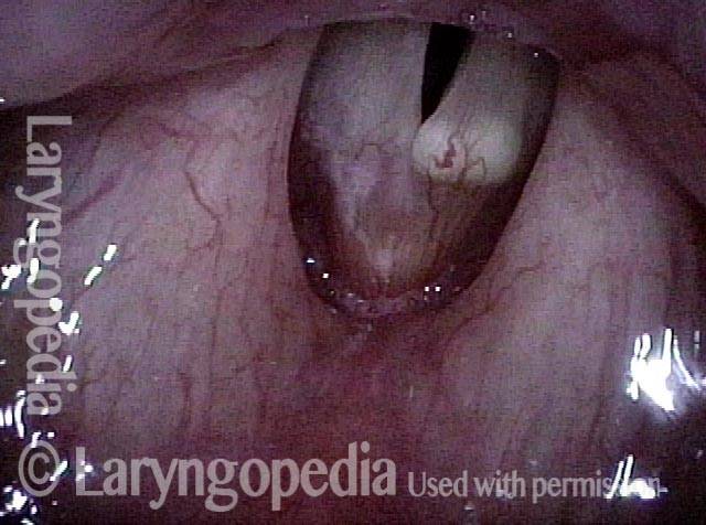
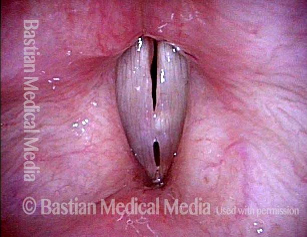
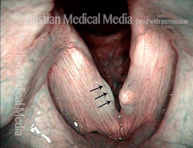
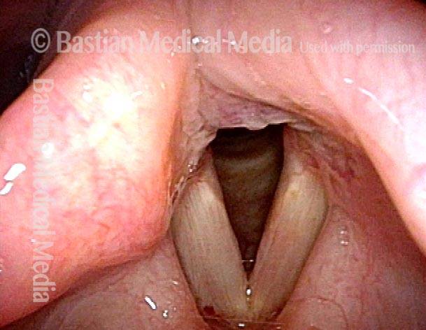
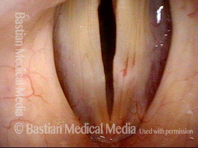
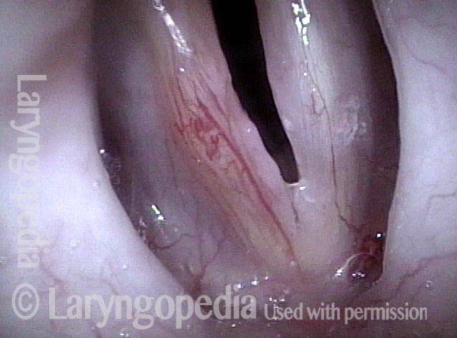
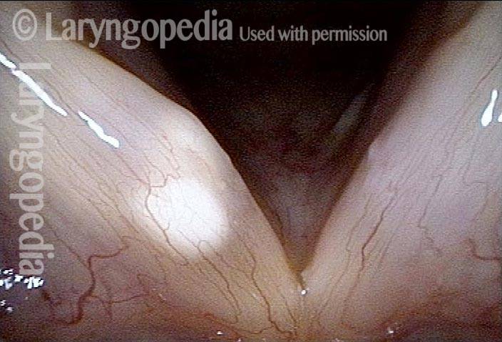
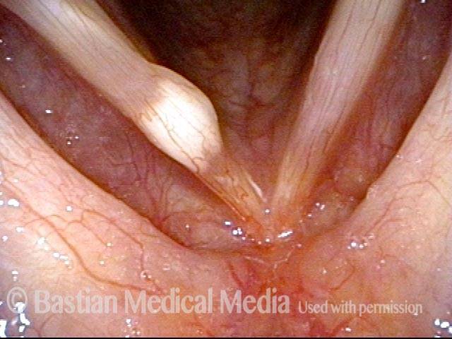
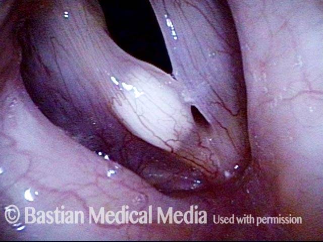
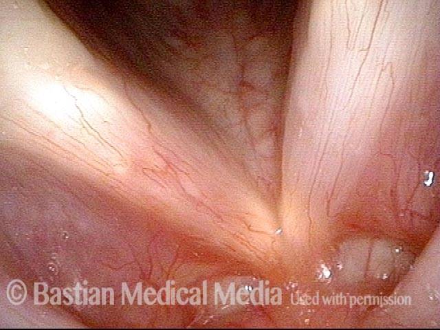
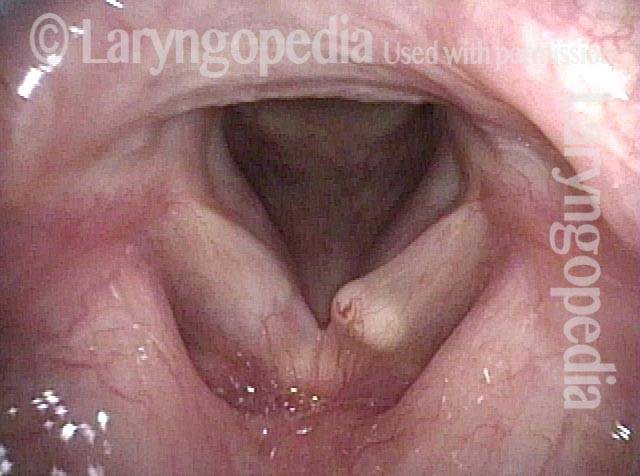
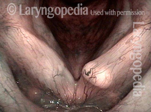
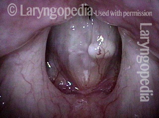
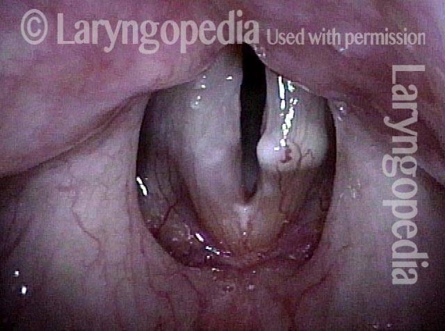
An epidermoid cyst of the vocal cord is generally most visible on the cord’s upper surface, and is whitish in color. In comparison with mucus retention cysts, epidermoid cysts project less from the cord, and when smaller, they can be quite subtle and easy to miss.

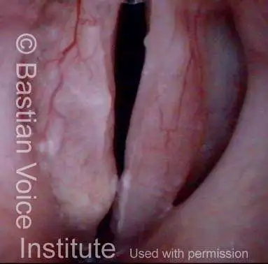
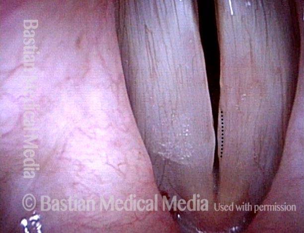
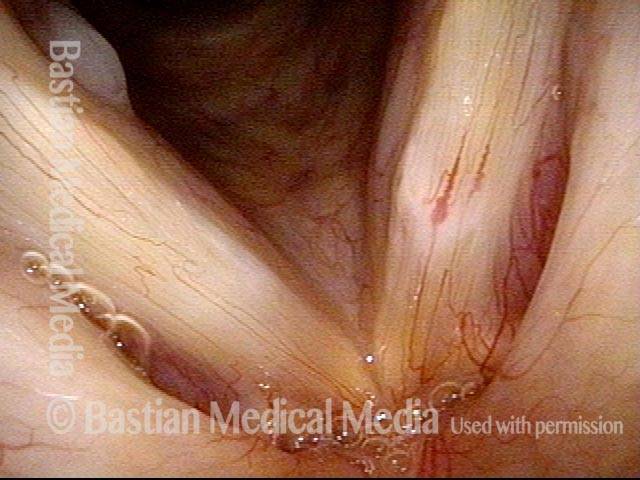
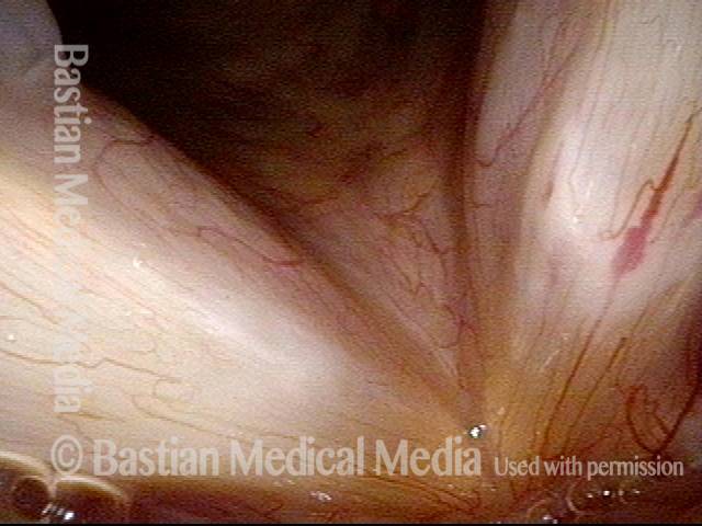
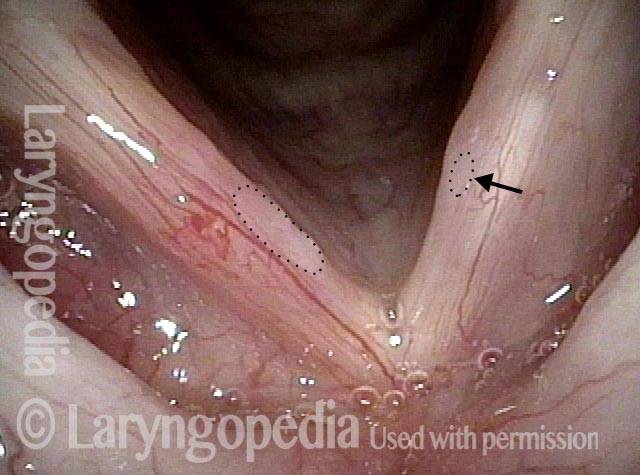
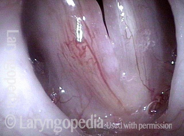
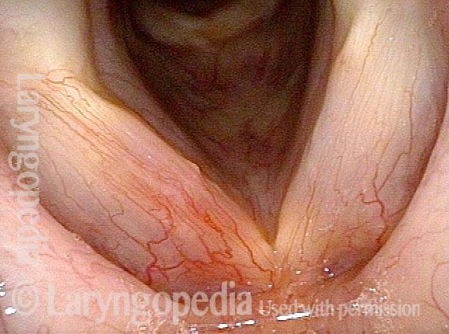
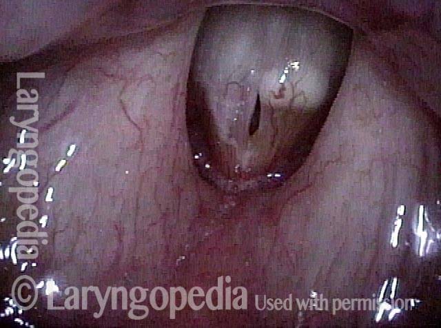
Sometimes an epidermoid cyst will have spontaneously ruptured but still retain some of the accumulated keratin (an open cyst); in this case, the cyst’s outline may be more subtle, and usually assumes an oval shape with the long axis oriented anteriorly and posteriorly. An open cyst may also produce a mottled appearance.

Una cisti epidermoide della corda vocale è generalmente più visibile sulla superficie superiore della corda ed è di colore biancastro. Rispetto alle cisti da ritenzione di muco, le cisti epidermoidi sporgono meno dal cordone e, quando sono più piccole, possono essere piuttosto sottili e facili da non notare.

A volte una cisti epidermoide si rompe spontaneamente ma conserva ancora parte della cheratina accumulata (una cisti aperta); in questo caso il contorno della cisti può essere più sottile, e solitamente assume una forma ovale con l’asse lungo orientato anteriormente e posteriormente. Una cisti aperta può anche produrre un aspetto chiazzato.

Trattamento per una cisti epidermoide

La logopedia è importante per i pazienti che esagerano con la voce, per aiutare a prevenire il verificarsi futuro di questa o altre lesioni e lesioni legate all’uso eccessivo della voce, ma non risolverà la cisti. È possibile eseguire un intervento chirurgico per rimuovere la cisti. Ciò richiede un’incisione e quindi la dissezione dell’intera sfera intatta della cisti.

L’intervento è tecnicamente molto più difficile della rimozione di noduli o polipi ed è più probabile che causi rigidità cronica della mucosa danneggiata. Tuttavia, i risultati possono essere molto buoni, soprattutto se la mucosa sovrastante è relativamente spessa.


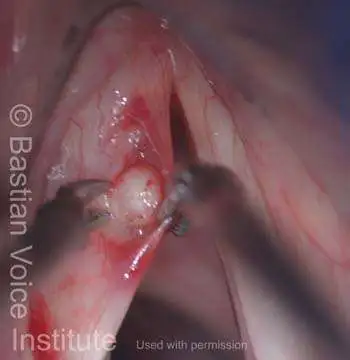
Rimozione della cisti

Visual Portfolio, Posts & Image Gallery for WordPress

Cyst removal (1 of 7)

Initial view with white sphere (cyst) shining through upper surface of left vocal fold.

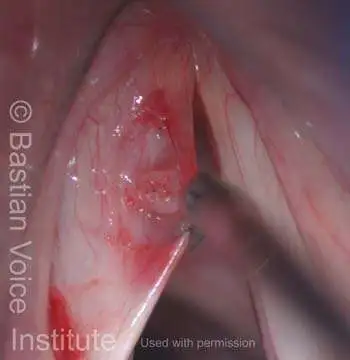
Cyst removal (2 of 7)

After injection of lidocaine for hydrodissection (Bouchayer).

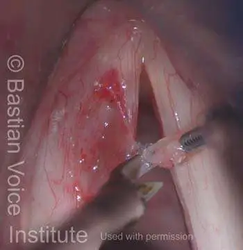
Cyst removal (3 of 7)

This incision must be ~75% longer than the diameter of the cyst. The most difficult parts of the dissection are typically adherence to the vocal ligament, and to the diaphanous overlying mucosal flap (held here in micro-alligator forceps).

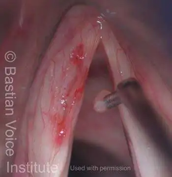
Cyst removal (4 of 7)

As is occasionally the case, the cyst has leaked some of its contents, but its outline is still clearly visible, allowing complete and precise removal.

Cyst removal (5 of 7)

Scissors are used to release the cyst from filmy attachments, precisely at its margin.

Cyst removal (6 of 7)

The flap is released and returned to its original position; the incipient sulcus is still seen.

Cyst removal (7 of 7)

Retraction of the flap reveals the gossamer nature of the still-intact overlying mucosa.

Cisti epidermoide

Visual Portfolio, Posts & Image Gallery for WordPress

Epidermoid cyst (1 of 2)

Right epidermoid cyst during breathing (Lab). Note whitish sphere not as prominent due to thicker overlying mucosa, vascularity and mucus, suggesting concurrent acid reflux.

Epidermoid cyst (2 of 2)

Same patient, during phonation, showing mismatch. In addition, right side very stiff and non-vibratile (Lab).

Esempio 2

Visual Portfolio, Posts & Image Gallery for WordPress

Teacher with longstanding hoarseness (1 of 4)

Elementary school teacher with longstanding hoarseness, told that she had a “nodule” on one vocal cord.

Epidermoid cyst and polypoid nodule seen (2 of 4)

Closer inspection, under narrow band light, shows that she has an epidermoid cyst of the left cord (right of photo) and a polypoid nodule with what looks like a small sulcus (arrows) of the right cord (left of photo).

Six days post-surgery, voice improved (3 of 4)

Six days after vocal cord microsurgery to address both lesions seen in photo 2, voice is already dramatically improved. This is a distant view under standard light.

Expected early postsurgical swelling (4 of 4)

Also six days postop, at close range under strobe light. As the cords approach phonation position, note the expected very early postsurgical swelling of the right cord (left of photo), and linear incision (tiny dots) through which the cyst was removed from the left cord (right of photo). No later postoperative photos are available.

Possibile cisti epidermoide aperta

Visual Portfolio, Posts & Image Gallery for WordPress

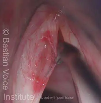
Capillary ectasia and white submucosal abnormality (1 of 3)

Left vocal cord (right of photo) has not only overlying capillary ectasia, but a white submucosal abnormality.

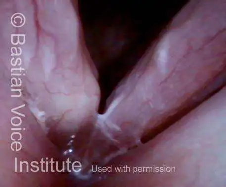
Prephonatory view, mtd (2 of 3)

Prephonatory instant shows some muscular tension dysphonia as well.

Open cyst (3 of 3)

Closer view. While an intact epidermoid cyst has a distinct outline, an open cyst develops a more mottled appearance. It may leak intermittently from a tiny dimple or sulcus which is sometimes seen at very close range.

Cisti e solco aperti nello stesso paziente

Visual Portfolio, Posts & Image Gallery for WordPress

Hoarse voice (1 of 4)

A young woman with a history of repeated loud cheering during athletic activities, to the point of hoarseness. She has a sulcus of the right cord (left of photo), and an open cyst of the left ( right of photo). Openings from sulcus and cyst are indicated by dotted lines.

Cyst + sulcus (2 of 4)

Narrow band light. The lateral lip of a sulcus is often bordered by a prominent capillary as seen here. An open cyst assumes an elliptical shape in the anteroposterior direction. It fails to empty completely because the opening draining it is smaller than the diameter of the cyst.

Closed phase (3 of 4)

Closed phase of vibration, strobe light.

Open phase (4 of 4)

Open phase of vibration.

Cisti epidermoide, prima e dopo la rimozione

Visual Portfolio, Posts & Image Gallery for WordPress

Epidermoid cyst (1 of 4)

A large and somewhat bi-lobed epidermoid cyst of the right vocal cord (left of photo). The cyst is white due to retained keratin. A mucus retention cyst tends instead to be either clear, or yellowish.

Phonatory view (2 of 4)

While making voice, the affected side does not vibrate.

Post microsurgery (3 of 4)

A week after microsurgical removal. The mucosa has been preserved, but capillaries are congested. The incision line on the upper surface of the cord is indicated by the subtle dotted line. Voice is already much improved.

4 months post surgery (4 of 4)

4 months after surgery, expected inflammatory change remains evident, but the incision line is no longer seen. The small white dot at the margin is mucus. Under strobe light, the right cord (left of photo) has regained partial ability to vibrate. Because its match with the left fold (right of photo) is precise, voice is surprisingly good, even at high pitch.

Esempio 2

Visual Portfolio, Posts & Image Gallery for WordPress

Epidermoid cyst (1 of 3)

Epidermoid cyst, right vocal cord. Note white submucosal mass predominately on upper surface of the cord, but with bilateral free margin elevation as well.

Epidermoid cyst (2 of 3)

Close view, initiation of phonation under strobe light.

Epidermoid cyst, removed (3 of 3)

After submucosal dissection and removal of cyst. Free margin swelling remains, as one cannot straighten the free margin (remove redundant mucosa stretched over the cyst) at the same time as cyst removal.

Cisti epidermoide bilobata e vibrazione scintillante

Visual Portfolio, Posts & Image Gallery for WordPress

Distant view (1 of 6)

This middle-aged coach has been grossly hoarse for many months. In this distant view, you can see a white submucosal mass of her left vocal cord (right of photo). It already appears to be an epidermoid cyst.

Closer view, narrow band light (2 of 6)

At close range under narrow band light one can observe a kind of bi-lobularity.

Closure under strobe light (3 of 6)

Under strobe light at the low pitch of B-flat 3 (233 Hz), closure is complete.

Open phase (4 of 6)

Open phase of vibration at the same pitch. Both cords open and the larynx creates just one pitch.

An octave above (5 of 6)

At an octave above, B-flat 4 (466 Hz), the left vocal cord (right of photo) no longer vibrates. The open phase of vibration includes only the anterior segment here.

“Shimmying” vocal cord (6 of 6)

The open phase a few frames later includes only the posterior segment. Clear sound is produced with this “shimmying” right vocal cord (left of photo) vibration. At other pitches the cord vibrates chaotically with two pitches.
Vocal nodules & other voice injuries YT Thumbnail
Riproduci video

Noduli e altre lesioni delle corde vocali: come si verificano e come possono essere trattati

Questo video spiega come si verificano i noduli e altre lesioni delle corde vocali: attraverso l’eccessiva vibrazione delle corde vocali, che si verifica con un uso eccessivo della voce. 

Dopo aver posto queste basi, il video prosegue esplorando il ruolo delle opzioni terapeutiche come la terapia vocale e la microchirurgia delle corde vocali.

Sommario

Subscribe
Notify of

0 Comments
Inline Feedbacks
View all comments
0
Click to see all comments.x