A mucus retention cyst forms when one of the mucus glands just below the vocal cord’s free margin becomes plugged. Mucus glands in this location secrete mucus in order to bathe and lubricate the vocal cords, but if a gland becomes obstructed, then the mucus it produces gets trapped and accumulates, leading to a mucus retention cyst. They typically occur without any correlation to vocal overuse, in contrast to epidermoid cysts as well as nodules and polyps.
Symptoms
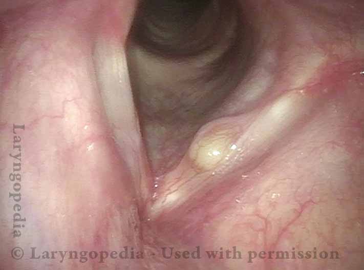
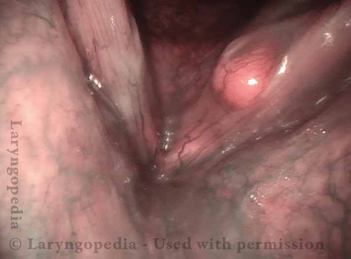
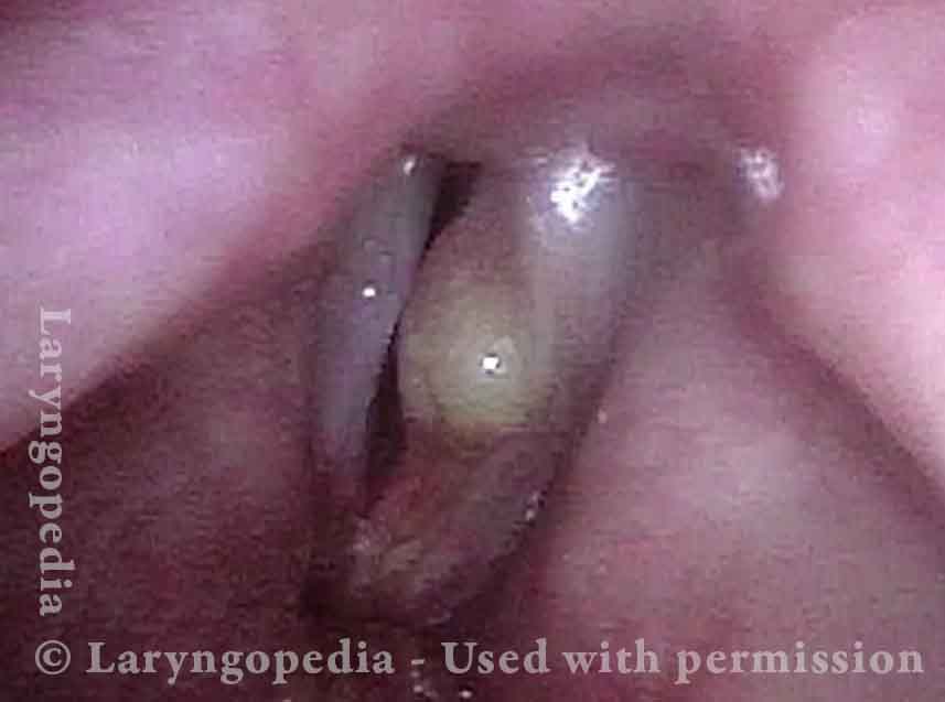
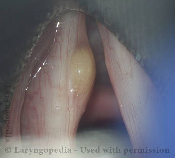
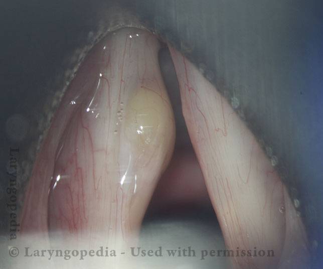
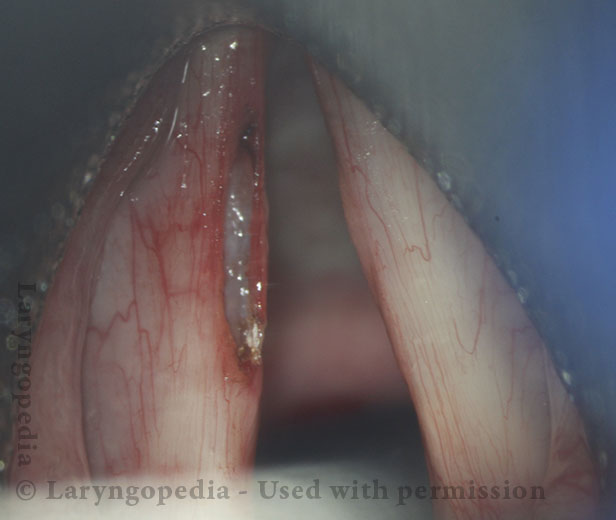
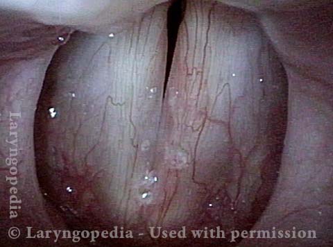
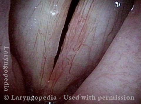
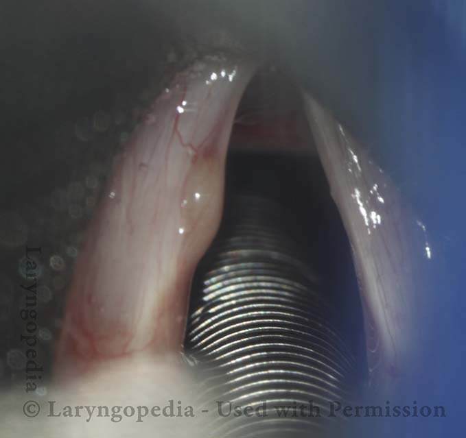
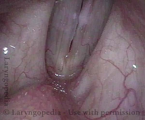
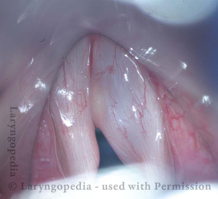
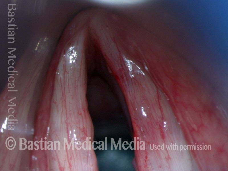
A mucus retention cyst can cause hoarseness, because it interferes with the normal vibrations of the vocal cords and the accuracy of their match with each other (see the videos below). The cyst is most often unilateral—that is, occurring on one cord but not the other. It appears as a bulge or deformation of the vocal cord’s free margin, and sometimes undersurface, and it may be yellowish in color.
Treatment
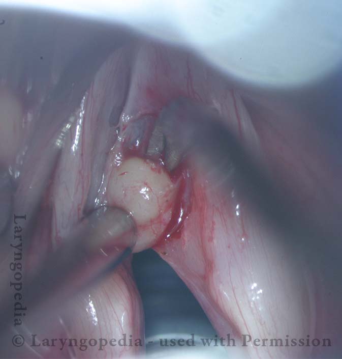
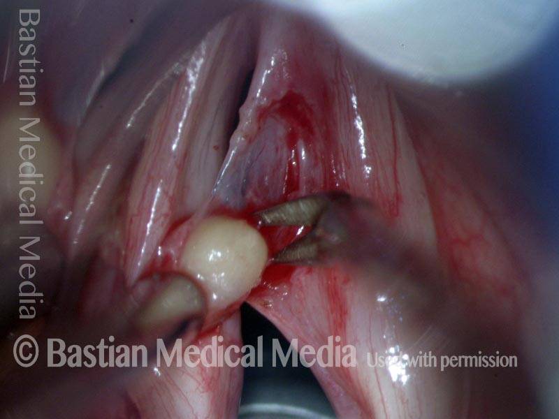
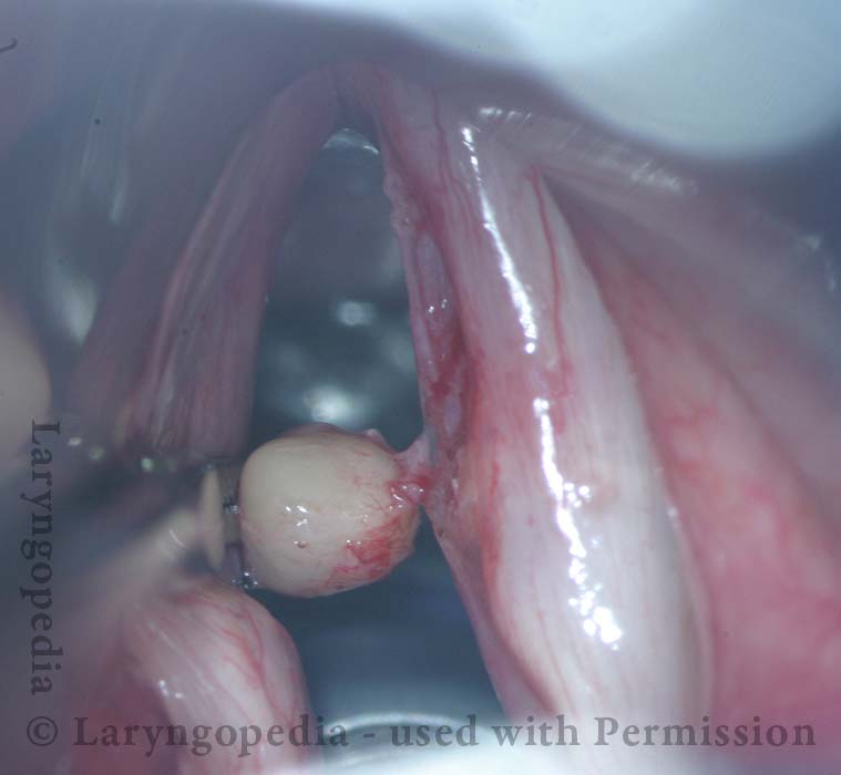
The cyst may be surgically removed, by creating a small incision on the vocal cord and then dissecting the cyst from the cord. Photos of the surgical process can be found below. Also, the two videos below show how removing this kind of cyst can improve the voice.
Excision of a Mucus Retention Cyst that Decompresses during Dissection
This man has had slight hoarseness for many years, possibly related to his self-described highly talkative and loud-spoken nature. His voice took a significant turn for the worse soon after a bout of mild laryngitis, and had remained extremely hoarse for many months.
An ENT doctor diagnosed a polyp of his left vocal cord. Instead, this represents a mucus retention cyst. Removal provided dramatic return of clear speaking voice. Seen below is preop, intra-operative sequence, and then the result at 3 months after surgery.
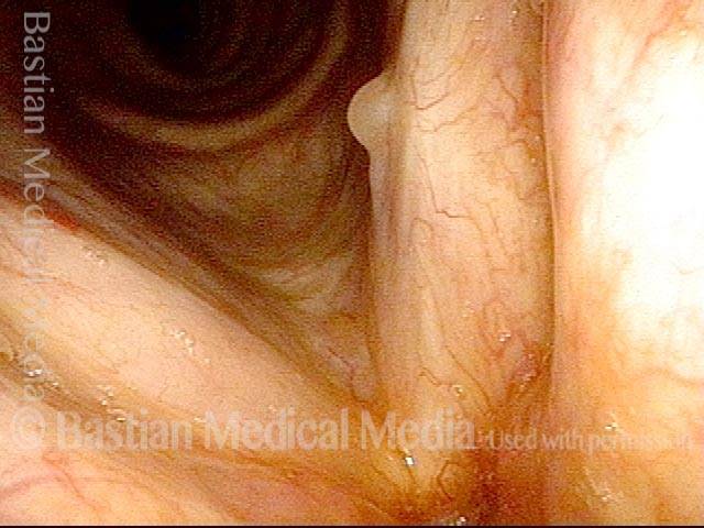
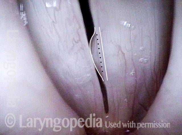
Mucus retention cyst (1 of 17)
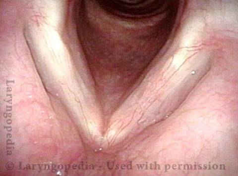
Closer look (2 of 17)
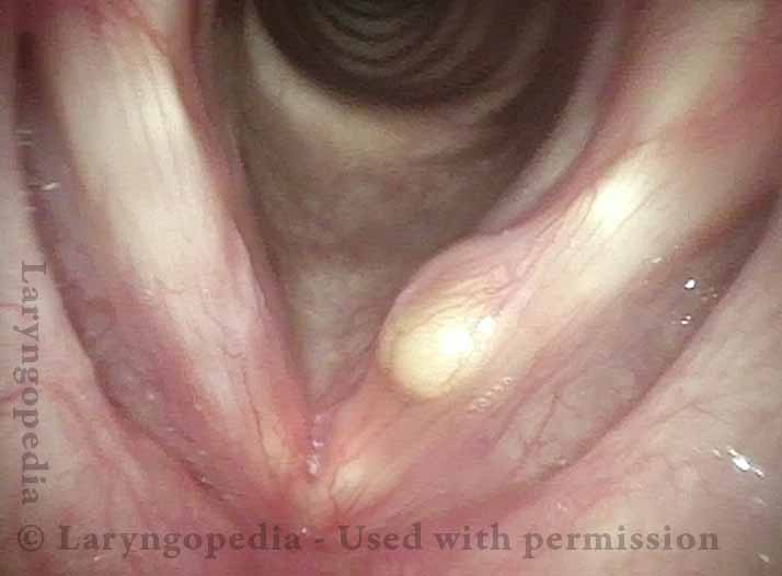
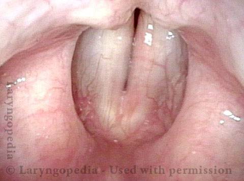
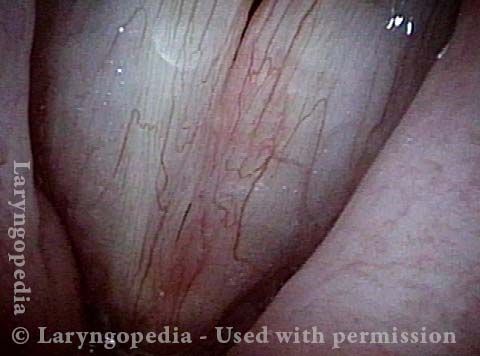
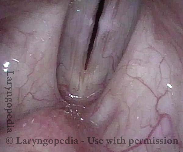
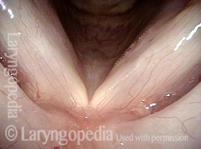
Cyst under narrow band light (3 of 17)
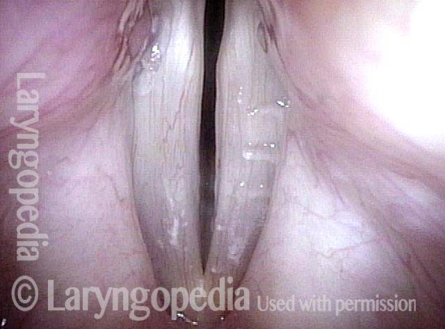
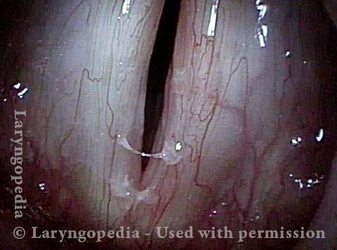
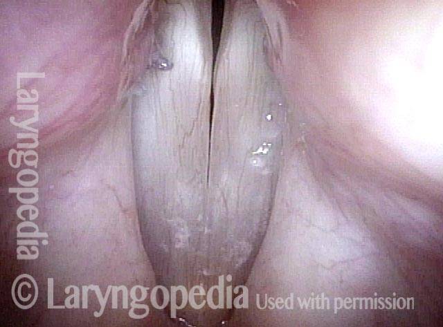
Cyst interferes with phonation (4 of 17)
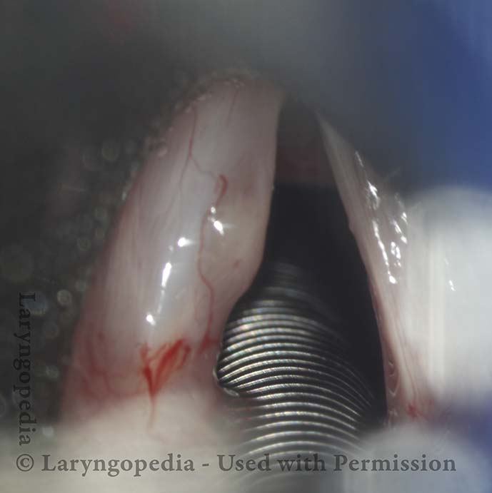
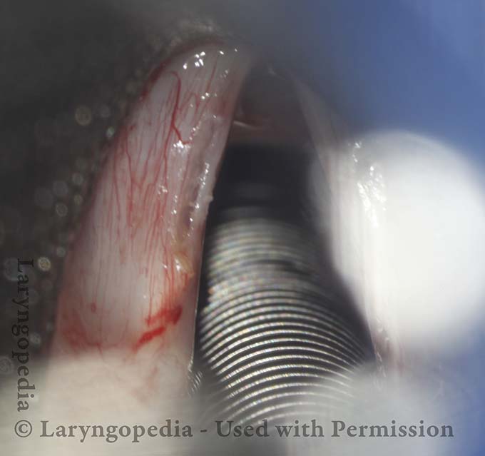
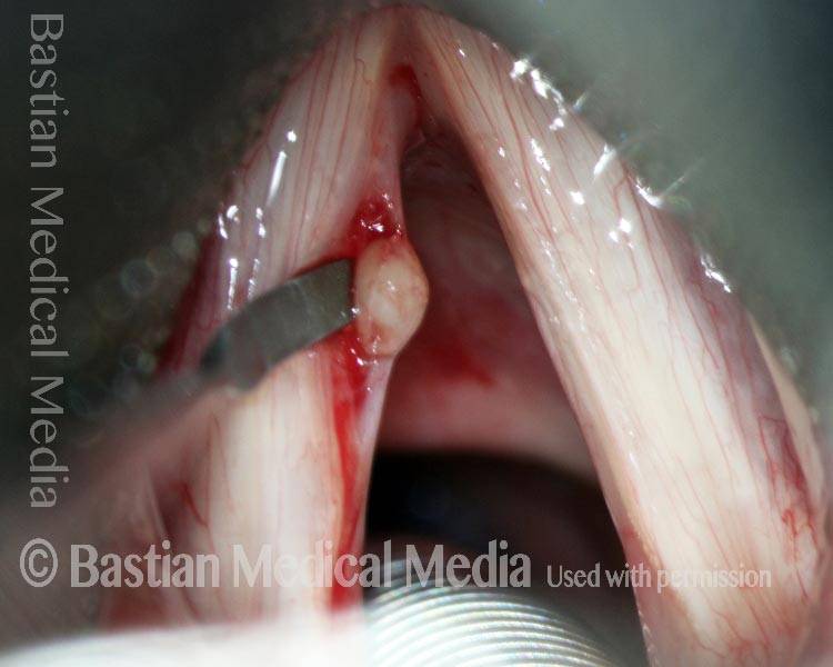
Surgical view (5 of 17)
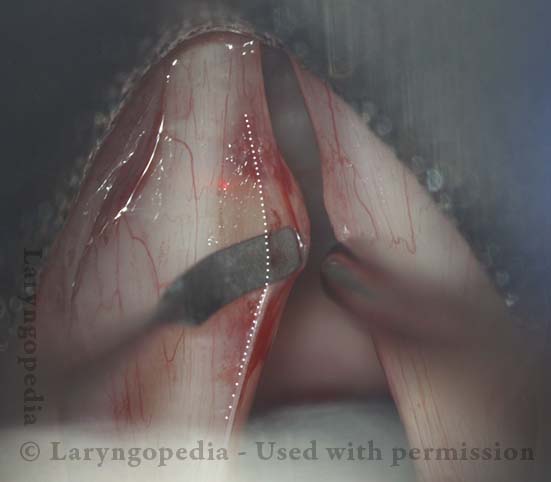
After infiltration (6 of 17)
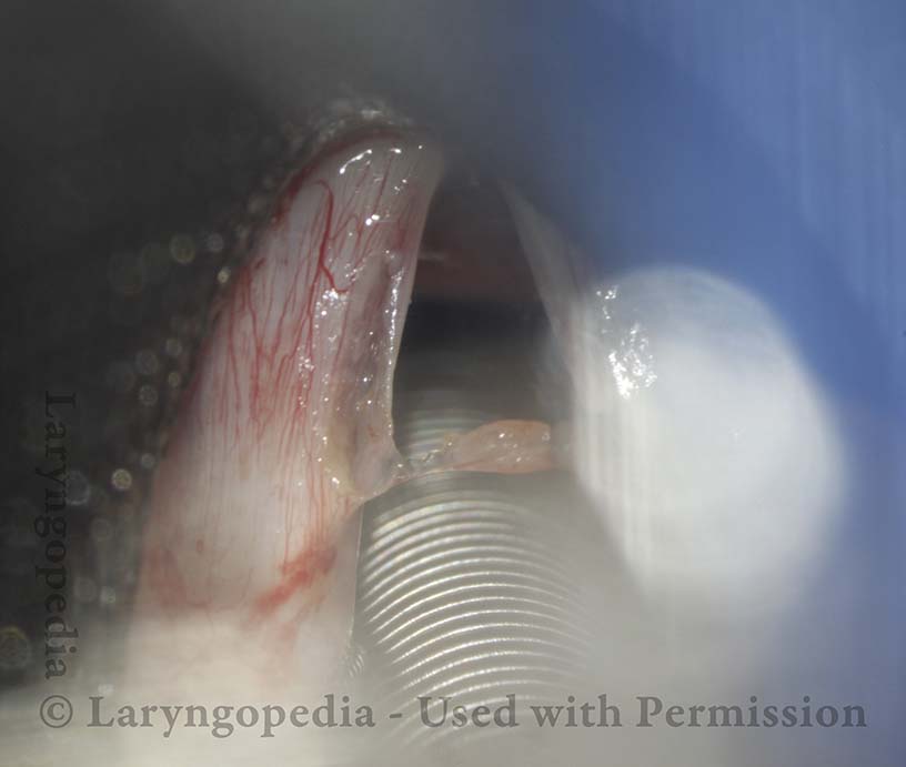
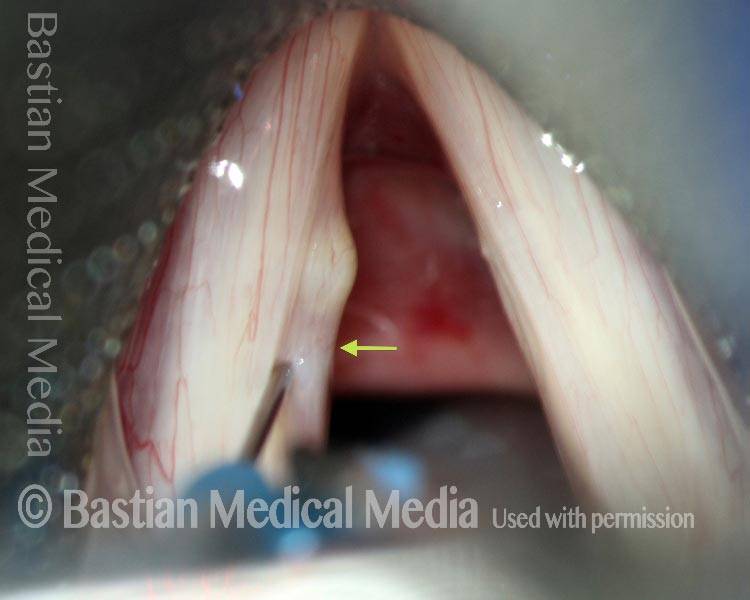
Lifting the mucosa (7 of 17)
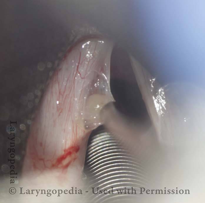
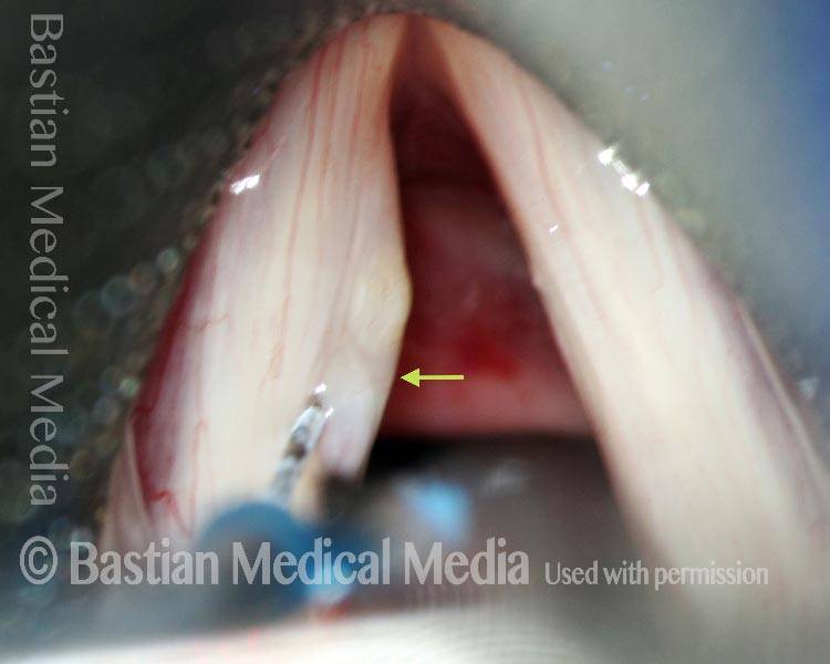
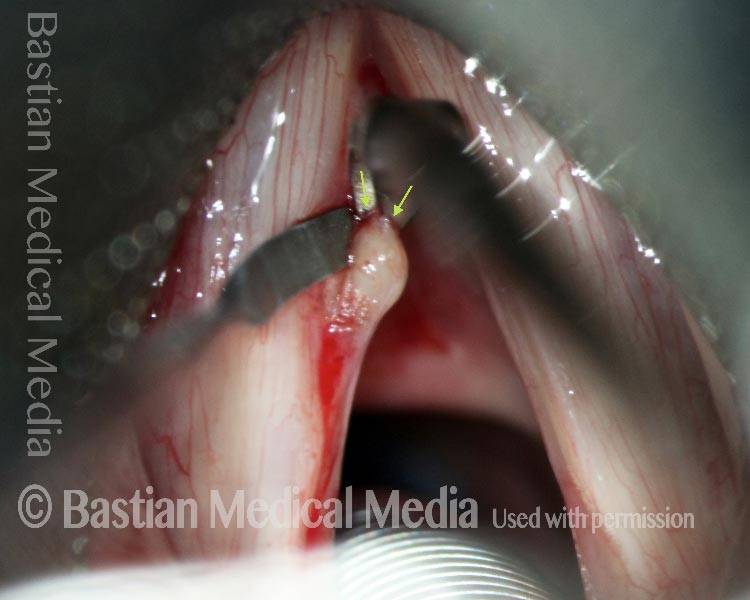
Dissecting cyst from deep attachments (8 of 17)
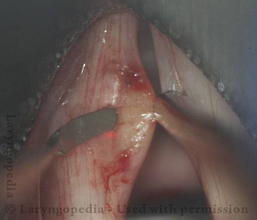
Cyst has ruptured and emptied (9 of 17)
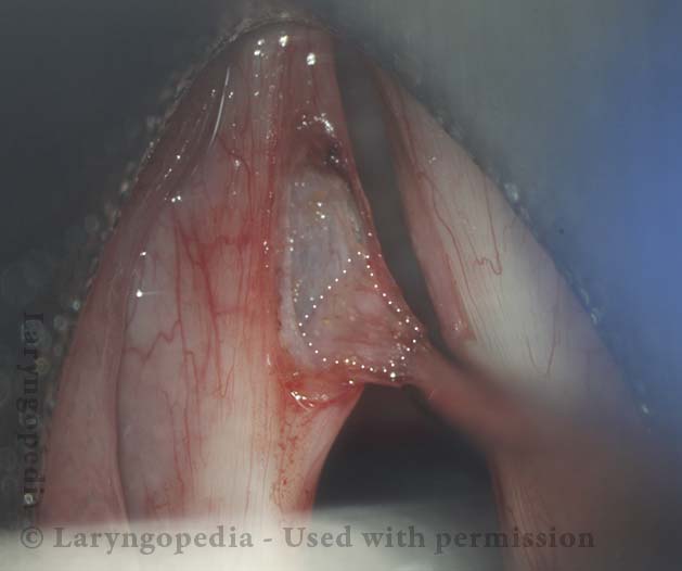
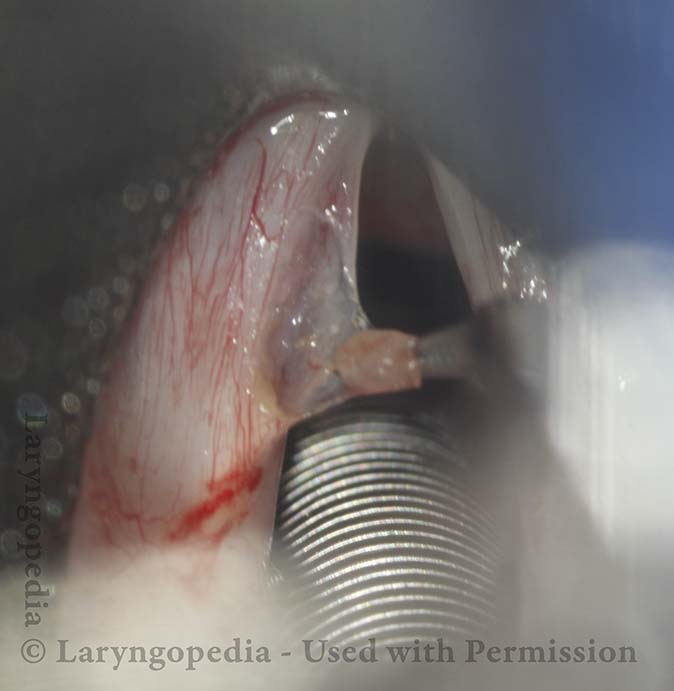
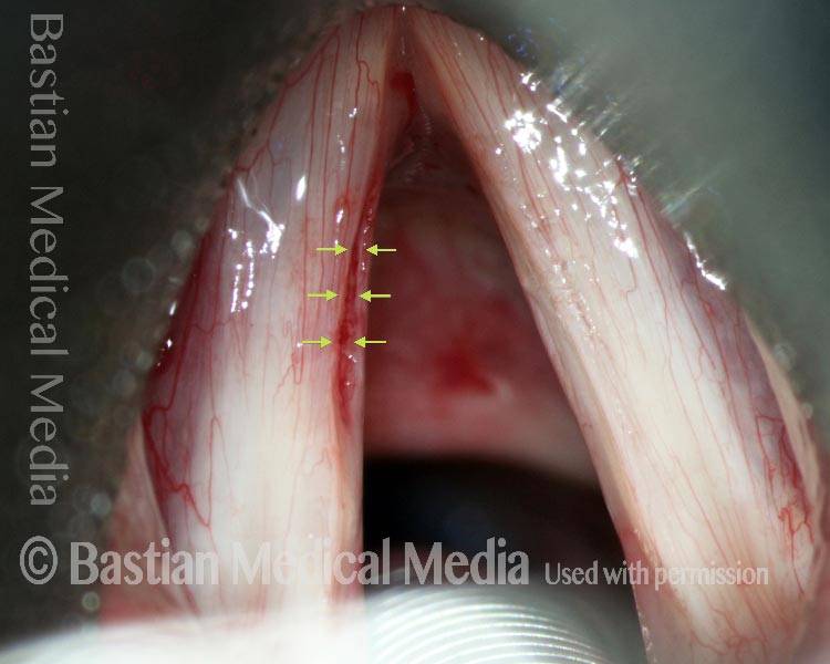
Dissection of empty sac (10 of 17)
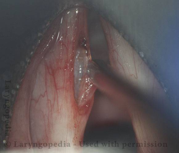
Flaps retract (11 of 17)
Voice is virtually normal at 3 months (12 of 17)
Evidence of vibration, standard light (13 of 17)
Open phase at E3 (14 of 17)
Closed phase at E3 (15 of 17)
Open phase at A4 (16 of 17)
Closed phase at A4 (17 of 17)
Removal of Mucus Retention Cyst, Still Intact
Mucus retention cyst (1 of 8)
Injection in Reinke’s space (2 of 8)
Incision begins (3 of 8)
Removal of cyst (4 of 8)
Removal nearly complete (5 of 8)
Cyst is removed (6 of 8)
Post-op (7 of 8)
Open Phase (8 of 8)
Removal of Mucus Retention Cyst
Mucus retention cyst (1 of 7)
Xylocaine prepares for removal (2 of 7)
Removal of Cyst (3 of 7)
Removal of cyst (4 of 7)
Removal of Cyst (5 of 7)
Voice immediately improves (6 of 7)
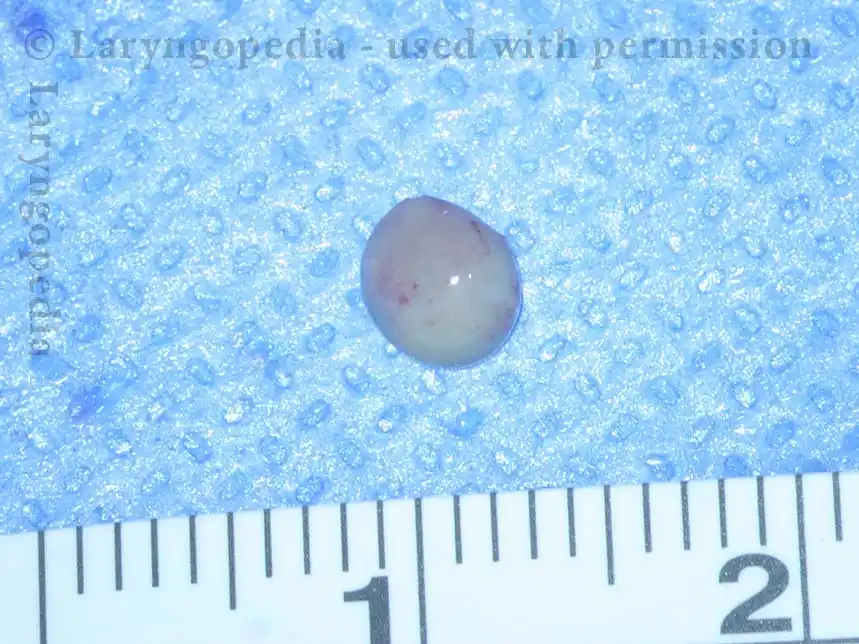
4mm Cyst (7 of 7)
Example 2
Injecting Xylocaine (1 of 5)
Hydrodissection effect (2 of 5)
Removal of cyst (3 of 5)
Removal of cyst (4 of 5)
Cyst is gone! (5 of 5)
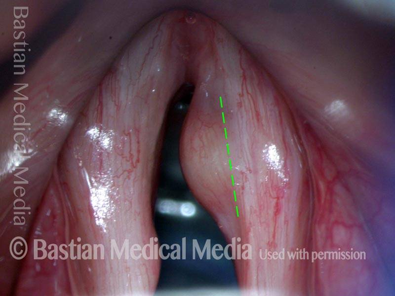
Mucus-Retention Cyst—not Polyp—Before and After Removal
Mucus-retention cyst (1 of 5)
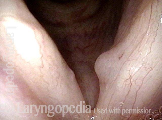
Below the margin (2 of 5)
One week post-op (3 of 5)
Better medializtion (4 of 5)
Better flexibility (5 of 5)