Post-swallow hypopharyngeal reflux refers to when, shortly after a person swallows, some swallowed material returns from below the esophageal entrance back up into the hypopharynx. This finding is an almost certain diagnostic indicator of cricopharyngeal dysfunction, usually with an associated Zenker’s diverticulum.
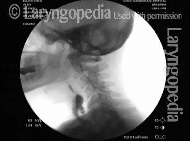
If this reflux occurs during a videoendoscopic swallowing study, the clinician will see that, though there may be little to no hypopharyngeal residue immediately after the swallow, a moment later some swallowed material (e.g., blue-stained applesauce or water) reappears and wells up in the post-arytenoid area and into the pyriform sinuses. If this reflux occurs during a videofluoroscopic swallowing study, the clinician will see barium remaining in the Zenker’s sac and, immediately after each swallow, moving back upward into the hypopharynx.
Suspect Zenker’s even if no Prior Radiographic Proof
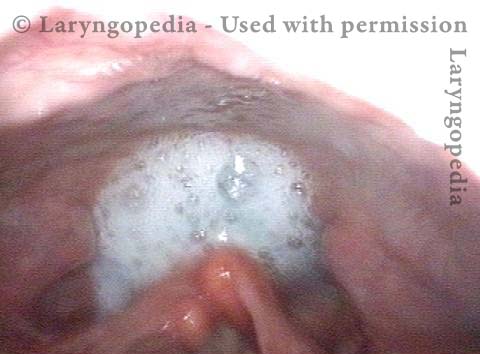
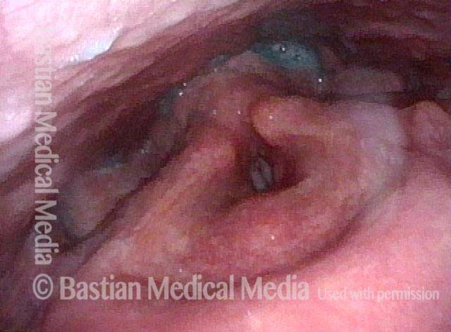
Dysphagia and possible Zenker's (1 of 5)
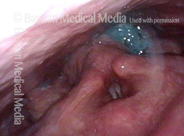
After a complete swallow (2 of 5)
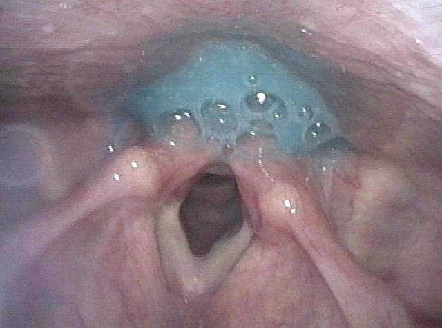
Additional material (3 of 5)
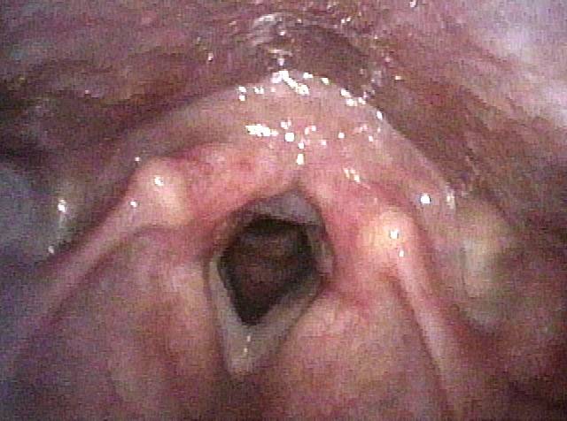
Hypopharyngeal residue (4 of 5)
Two seconds later (5 of 5)
Post-swallow Hypopharyngeal Reflux (Zenker’s Diverticulum): VESS vs. VFSS
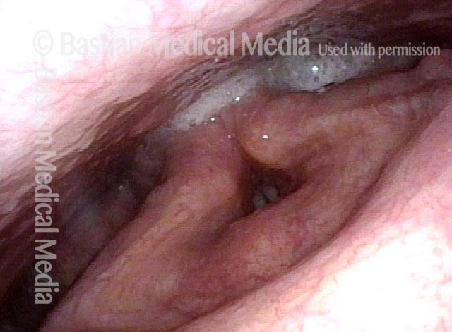
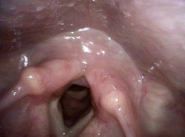
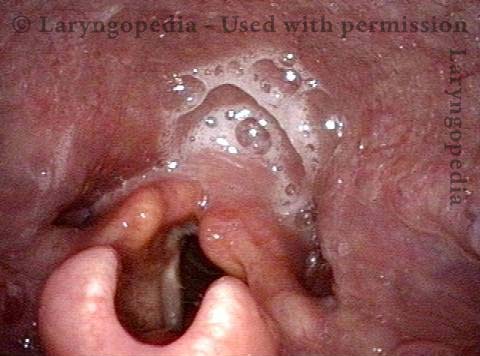
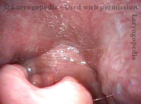
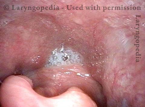
VESS (1 of 7)
Reflux (2 of 7)
Post-swallow (3 of 7)
Reflux of cracker (4 of 7)
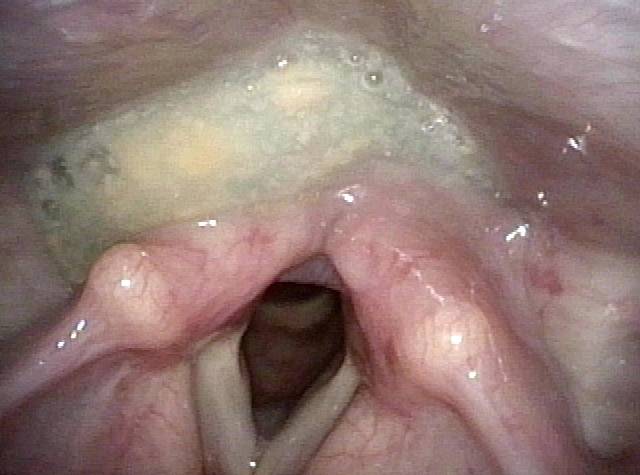
VFSS (5 of 7)
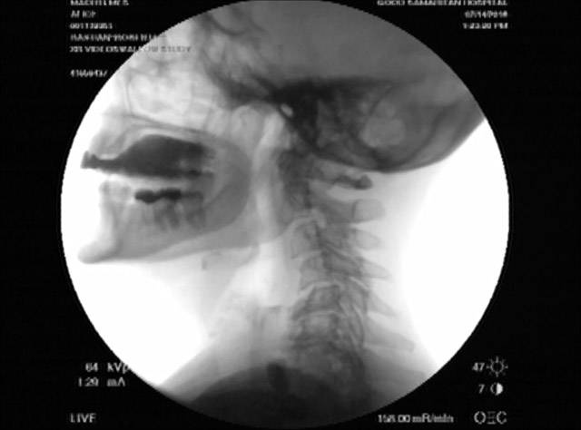
X-Ray (6 of 7)
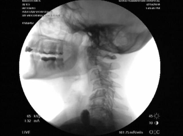
Continued reflux (7 of 7)
Post-swallow Return of Saliva into the Hypopharynx
This man began to experience solid food lodgment a few years earlier, along with gurgling noises every time he swallows. Even on an initial examination, Zenker’s diverticulum can be strongly suspected based on the findings of the videoendoscopic swallow study (VESS). Upon review of a videofluoroscopic swallow study (VFSS), a Zenker’s diverticulum is indeed the explanation for the findings below, which went away completely (return of saliva, gurgling noises, and food lodgment) after myotomy.
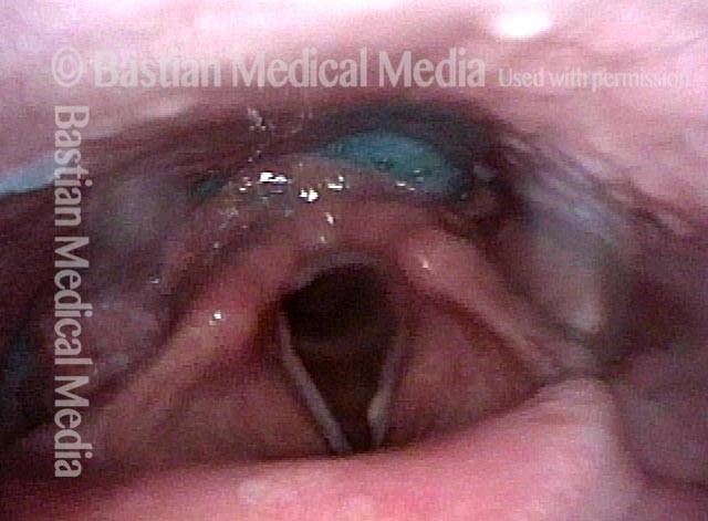
Pooling of saliva (1 of 4)
Saliva disappears after swallow (2 of 2)
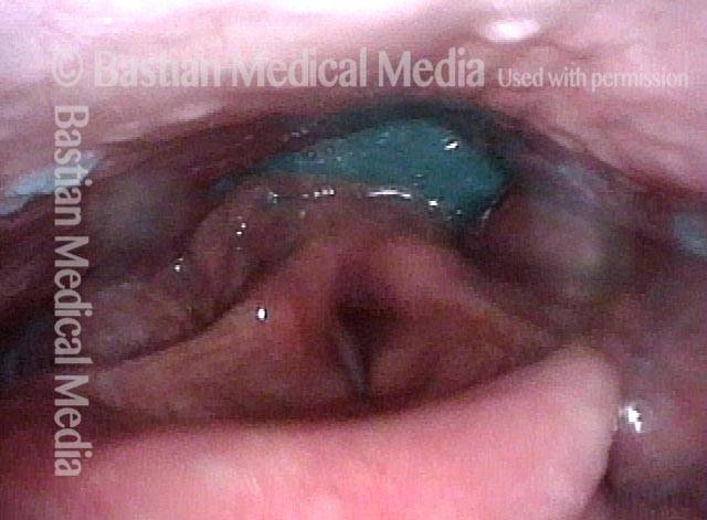
Saliva begins to return (3 of 3)
Saliva is ejected upwards from Zenker’s sac (4 of 4)