Photo Essay of Spindle Cell Carcinoma
This older man developed unexplained hoarseness. The original removal pathology result described only a “polyp.” Elsewhere, removal was complete, but recurrence is shown here that remarkably resembles the original tumor removed. Notice the mucosa-covered polypoid appearance though the tissue itself is very firm.
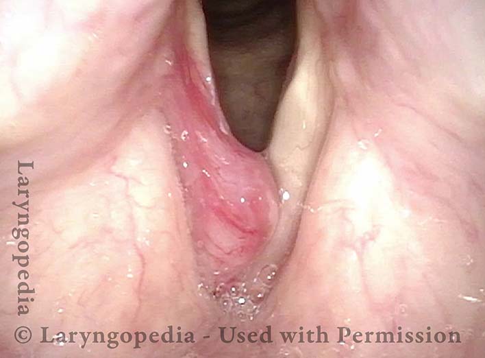
Tumor on Vocal Cord (R) (1 of 8)
From a distance, the rounded, mucosa-covered lesion of the right cord is seen.
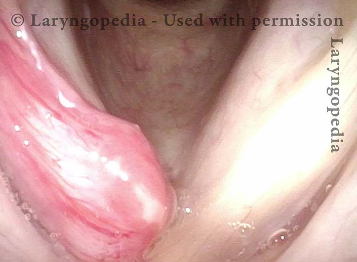
Tumor on Vocal Cord (R) (2 of 8)
At closer range, one can see that the left vocal cord is not involved.
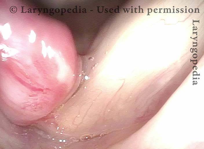
Anterior of tumor (3 of 8)
Better view of the anterior limit of the lesion.
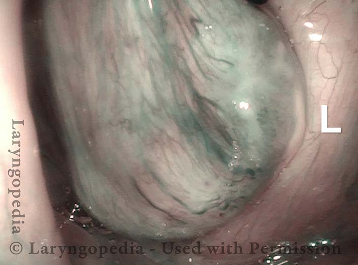
Capillaries over tumor (4 of 8)
Under narrow band light, abundant capillaries course through covering mucosa. “L” indicates the normal left vocal cord.
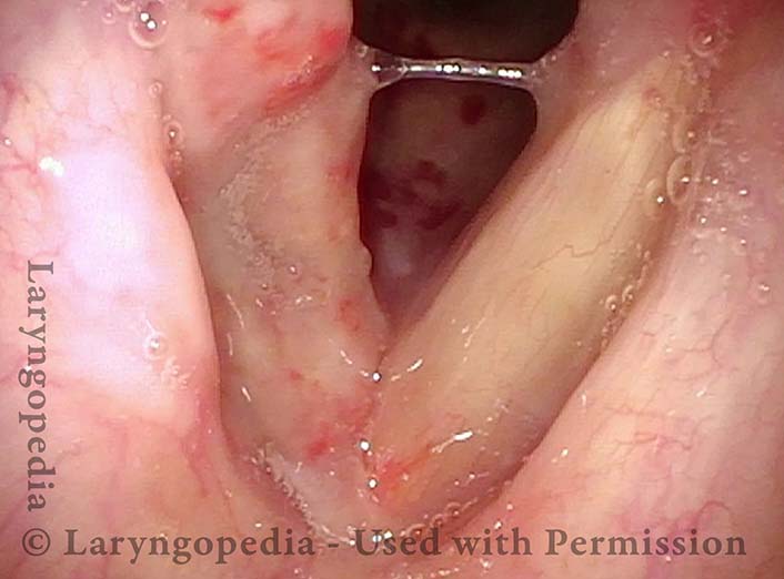
Tumor Removed (5 of 8)
One day after surgical removal. The excision was “definitive” but followed what seemed like a well-circumscribed lesion into muscle with a narrow margin in order to preserve normal tissue.
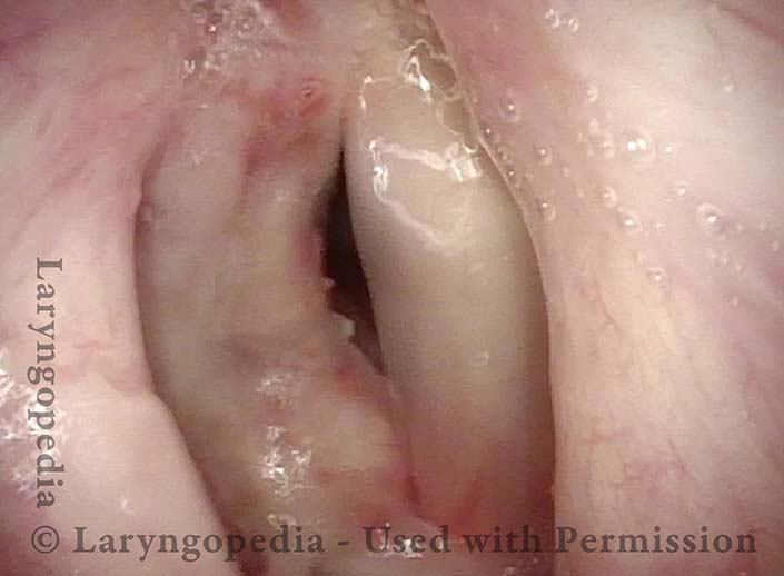
Functional but weak voice (6 of 8)
Voice is functional but weak, due to tissue loss and resultant phonatory gap. With healing, voice is expected to improve.
Granuloma or Tumor? (7 of 8)
At one month postop, healing is well underway. Granulomas are often seen during healing when excision went into vocal cord muscle. The question: is this a resolving granuloma, or recurrent tumor? This small lesion will be removed for testing, if still present at the next postoperative visit.
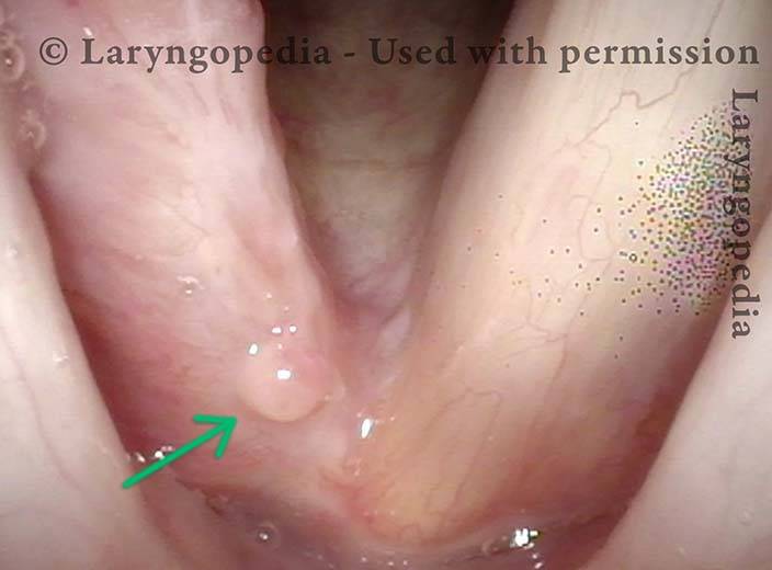
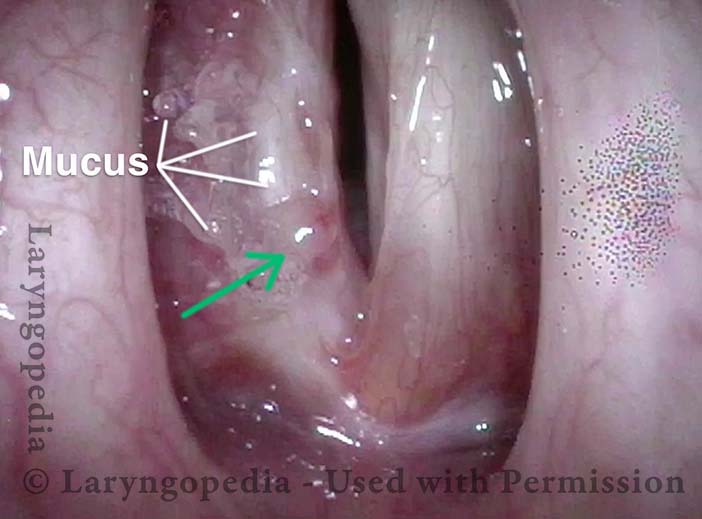
Cords match during phonation (8 of 8)
During phonation, vocal cords match better than immediately postoperatively. Arrow again indicates the “granuloma/recurrence.” The lines lead to mucus.