An encyclopedia about voice, swallowing, airway, coughing, & other head + neck disorders.

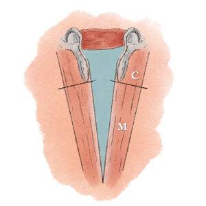
Illustration of the membranous glottis and cartilaginous glottis

Cartilaginous glottis (C) is the posterior one-third of the vocal cord’s visible length and also, during breathing, the space between this segment of both cords. It is inhabited by the arytenoid cartilage and covered by a relatively thin layer of perichondrium and, on top of that, a layer of mucosa.

It is here that contact granulomas occur, on the cord’s medial surface. The other two-thirds of each vocal cord’s visible length is called the membranous glottis (M).
Visual Portfolio, Posts & Image Gallery for WordPress

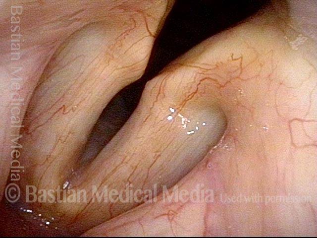
Cartilaginous glottis (1 of 1)

Vocal cords approaching adducted (phonatory) position. Note that the posterior 1/3 of the vocal cord is “inhabited” by the arytenoid cartilage, with vocal process turned slightly medially. The cartilaginous glottis appears to make up more than the posterior 1/3 of the cord due to optical distortion from the wide-angle lens used in the tip of the endoscope.

Unusual Posterior and Transglottic Epicenter for Larynx Cancer

Visual Portfolio, Posts & Image Gallery for WordPress

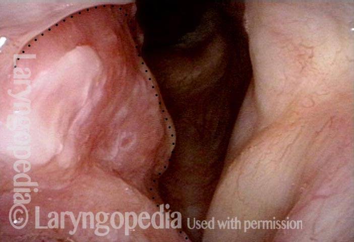
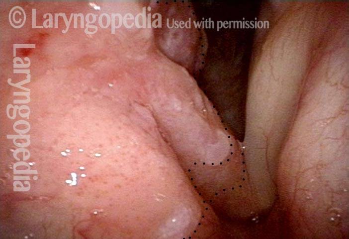
Tumor (1 of 4)

View under standard light shows normal left vocal cord (right of photo) and tumor on right (left of photo). Main bulk is posterior; the cord is mobile, yet (unusually) the tumor is transglottic.

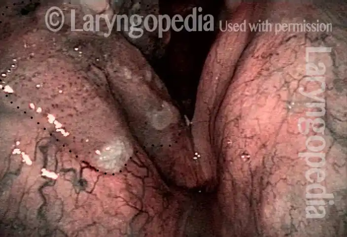
Accentuation of the vascularity (2 of 4)

Slightly more distant view under narrow band light; accentuation of the vascularity makes the tumor even more easily seen.

Posterior commissure (3 of 4)

Magnified posterior commissure view shows that the main bulk of tumor involves the cartilaginous glottis. This is highly unusual.

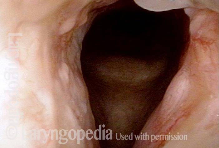
A year later (4 of 4)

Laser excision was satisfying except that the deep margin was the cricoid cartilage and therefore radiation therapy followed laser excision for added "safety." Laser excision and radiotherapy were 9 years prior to this posting, at which point he remained free of disease.