Incapacità di Ruttare
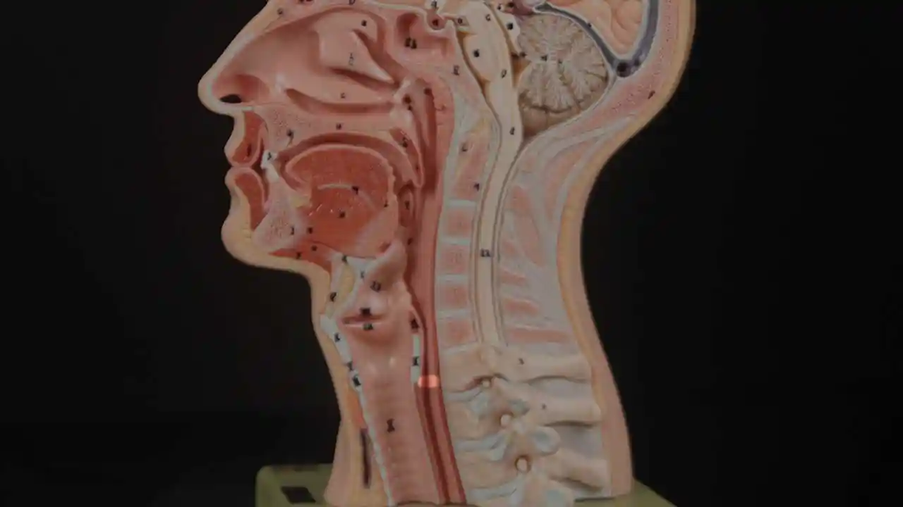
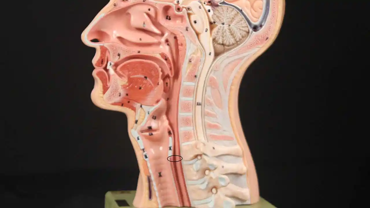
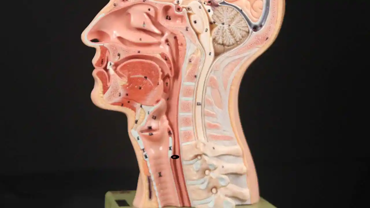
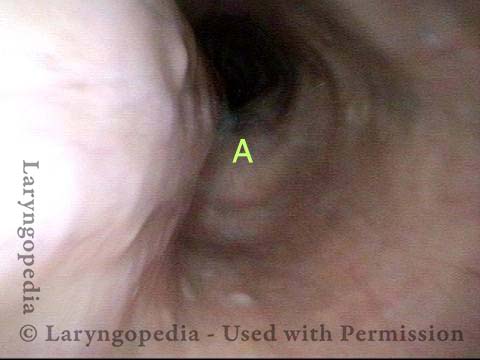
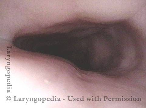
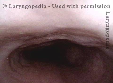
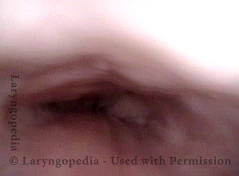
L’incapacità di ruttare si verifica quando lo sfintere esofageo superiore (muscolo cricofaringeo) non può rilassarsi per rilasciare la “bolla” d’aria. Lo sfintere è una valvola muscolare che circonda l’estremità superiore dell’esofago appena sotto l’estremità inferiore del passaggio della gola. Se guardi di fronte il collo di una persona, è appena sotto il “pomo di Adamo / Eva”, direttamente dietro la cartilagine cricoidea.
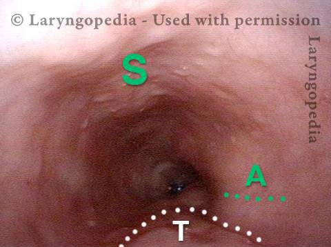
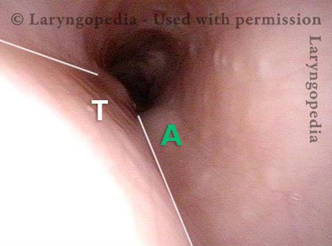
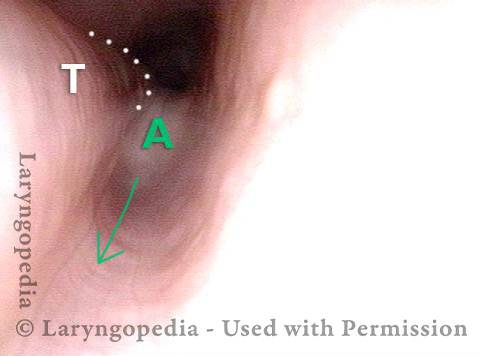
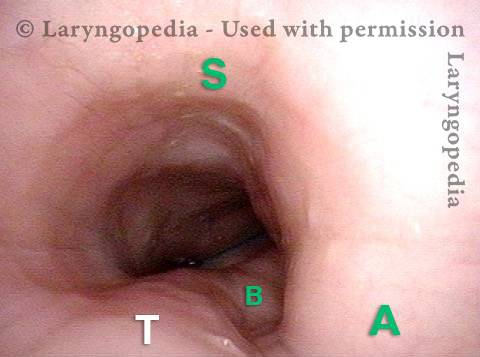
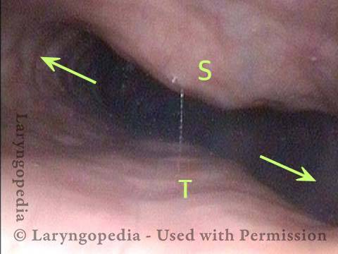
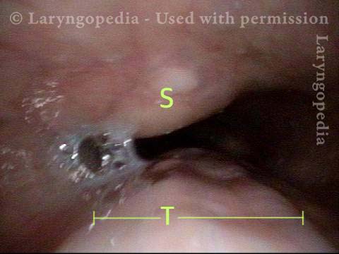
Se ti interessa vederlo su un modello, guarda le foto qui sotto. Il muscolo sfintere si rilassa per circa un secondo ogni volta che ingeriamo saliva, cibo o bevande. Tutto il resto del tempo è contratto. Ogni volta che una persona rutta, lo stesso sfintere deve lasciarsi andare per una frazione di secondo affinché l’aria in eccesso fuoriesca verso l’alto. In altre parole, così come è necessario che lo sfintere “lasci andare” per far entrare cibo e bevande verso il basso nel normale atto di deglutizione, è anche necessario che lo sfintere sia in grado di “lasciarsi andare” per rilasciare aria verso l’alto per eruttare. Il nome formale di questo disturbo è disfunzione cricofaringea retrograda (R-CPD).
Le persone che non possono rilasciare aria verso l’alto sono infelici. Possono sentire la “bolla” seduta dal collo medio a basso senza un posto dove andare. Oppure sperimentano gorgoglii quando l’aria sale dall’esofago solo per scoprire che la via di fuga è bloccata da uno sfintere non rilassante. È come se il muscolo dell’esofago si agitasse e si contraesse continuamente, senza successo.
La persona vuole e ha bisogno di ruttare, ma continua a sperimentare questa incapacità di ruttare. A volte questo può anche essere doloroso. Queste persone spesso sperimentano pressione toracica o gonfiore addominale e persino distensione addominale. Anche la flatulenza è grave nella maggior parte delle persone con R-CPD. Altri sintomi meno universali sono nausea dopo aver mangiato, singhiozzo doloroso, ipersalivazione o sensazione di difficoltà a respirare con sforzo quando “pieno d’aria”. Molte persone con R-CPD sono state sottoposte a test approfonditi e prove di trattamento senza benefici. R-CPD riduce la qualità della vita ed è spesso socialmente dirompente e provoca ansia. Le diagnosi comuni (errate) sono “reflusso acido” e “sindrome dell’intestino irritabile” e quindi i trattamenti per queste condizioni non alleviano i sintomi in modo significativo.
Approcci per il trattamento dell’incapacità di ruttare:
Per le persone che corrispondono alla sindrome di:
1) Incapacità di ruttare
2) Rumori gorgoglianti
3) Pressione toracica/addominale e gonfiore
4) Flatulenza
Ecco il modo più efficace per procedere: in primo luogo, una consultazione per determinare se i criteri per la diagnosi di R-CPD sono soddisfatti o meno. Successivamente, un semplice studio della deglutizione videoendoscopico in studio che incorpora un esame neurologico dei muscoli della lingua, della faringe (gola) e della laringe e spesso include una mini-esofagoscopia. Ciò stabilisce che lo sfintere funziona normalmente in una direzione di deglutizione in avanti (anterograda), ma non in modo inverso (retrogrado) con rutti o rigurgiti. Insieme ai sintomi sopra descritti, questa semplice consultazione ambulatoriale e valutazione della deglutizione stabilisce la diagnosi di disfunzione cricofaringea retrograda (non rilassamento).
Il secondo passo è inserire Botox nel muscolo sfintere malfunzionante. L’effetto desiderato di Botox nel muscolo è quello di indebolirlo per almeno diversi mesi. La persona ha quindi molte settimane per verificare che il problema sia risolto o almeno ridotto al minimo.
L’iniezione di Botox potrebbe potenzialmente essere eseguita in un ufficio, ma consigliamo la prima volta (almeno) di posizionarlo durante un’anestesia generale molto breve in una sala operatoria ambulatoriale. Questo perché la prima volta è importante rispondere alla domanda in modo definitivo, cioè che l’incapacità dello sfintere di rilassarsi quando viene presentato con una bolla d’aria dal basso, è il problema. Inoltre, sulla base di un’esperienza con oltre 1.020 pazienti a luglio 2022, una singola iniezione sembra “allenare” il paziente a ruttare. Circa l’80% dei pazienti ha mantenuto la capacità di ruttare molto tempo dopo che l’effetto del Botox si è dissipato. Cioè, da 6 mesi trascorsi dal momento dell’iniezione.
Patients treated for R-CPD as just described should experience dramatic relief of their symptoms. And to repeat, our experience in treating more than 890 patients (and counting) suggests that this single Botox injection allows the system to “reset” and the person may never lose his or her ability to burp. Of course, if the problem returns, the individual could elect to pursue additional Botox treatments, or might even elect to undergo endoscopic laser cricopharyngeus myotomy. To learn more about this condition, see Dr. Bastian’s description of his experience with the first 51 of his much larger caseload.
I pazienti trattati per R-CPD come appena descritto dovrebbero provare un sollievo drammatico dei loro sintomi. E per ripetere, la nostra esperienza nel trattamento di più di 1.020 pazienti (e il conteggio) suggerisce che questa singola iniezione di Botox consente al sistema di “ripristinarsi” e la persona potrebbe non perdere mai la sua capacità di ruttare. Naturalmente, se il problema si ripresenta, l’individuo potrebbe scegliere di perseguire ulteriori trattamenti di Botox, o potrebbe anche scegliere di sottoporsi a miotomia cricofaringea laser endoscopica. Per saperne di più su questa condizione, vedere la descrizione del Dr. Bastian della sua esperienza con i primi 51 dei suoi casi molto più grandi.
Sommario

Pannello R-CPD
Un gruppo di discussione di esperti sulla disfunzione cricofaringea retrograda (RCPD AKA “No Burp Syndrome”) con domande e risposte dai leader del settore.
Webinar R-CPD (senza rutto).
Webinar live R-CPD (senza rutto) ospitato dal Dr. Bastian il 26 luglio 2022 alle 18:00. CST
R-CPD Domande e risposte—Parte I
Il Dr. Bastian risponde a un elenco di domande inviate dai nostri partecipanti al webinar R-CPD.
Share this article
Dov'è il Muscolo Cricofaringeo?
Cricopharyngeus Muscle (1 of 3)
Cricopharyngeus Muscle (1 of 3)
Open Cricopharyngeus Muscle (2 of 3)
Open Cricopharyngeus Muscle (2 of 3)
Closed (3 of 3)
Closed (3 of 3)
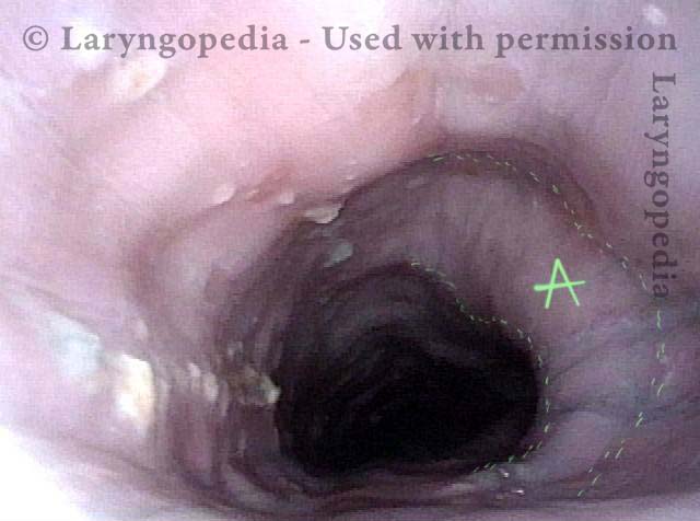
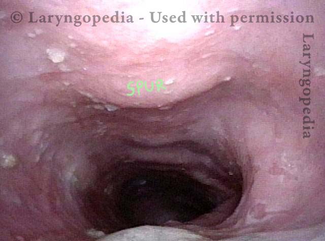
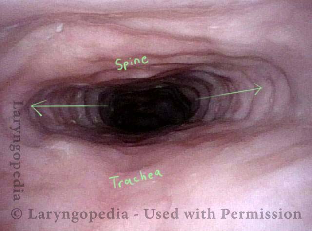
Reperti esofagei
Aortic shelf (1 of 3)
Aortic shelf (1 of 3)
Bony spur emerges due to stretched esophagus (2 of 3)
Bony spur emerges due to stretched esophagus (2 of 3)
Stretched esophagus due to unburpable air (3 of 3)
Stretched esophagus due to unburpable air (3 of 3)
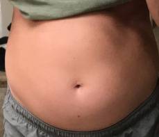
Distensione addominale di R-CPD
Gastric Air Bubble (1 of 3)
Gastric Air Bubble (1 of 3)
Bloated Abdomen (2 of 3)
Bloated Abdomen (2 of 3)
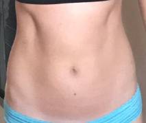
Non-bloated Abdomen (3 of 3)
Non-bloated Abdomen (3 of 3)
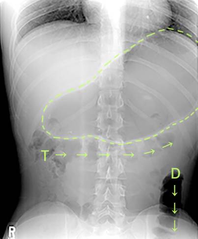
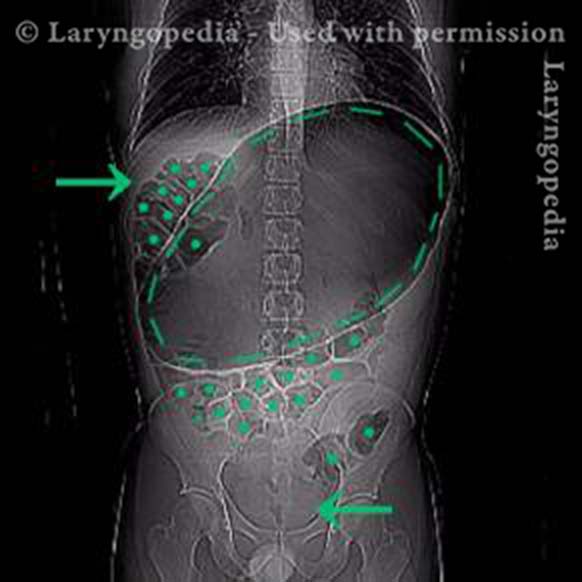
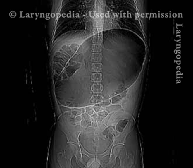
Una rara "crisi addominale" dovuta a R-CPD (incapacità di ruttare)
X-Ray of Abdominal Bloating (1 of 2)
X-Ray of Abdominal Bloating (1 of 2)
Impossibile ruttare: progressione del gonfiore e della distensione addominale—Un ciclo quotidiano per molti con R-CPD
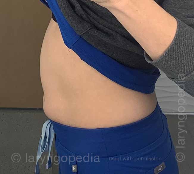
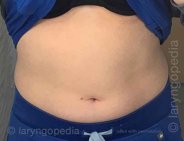
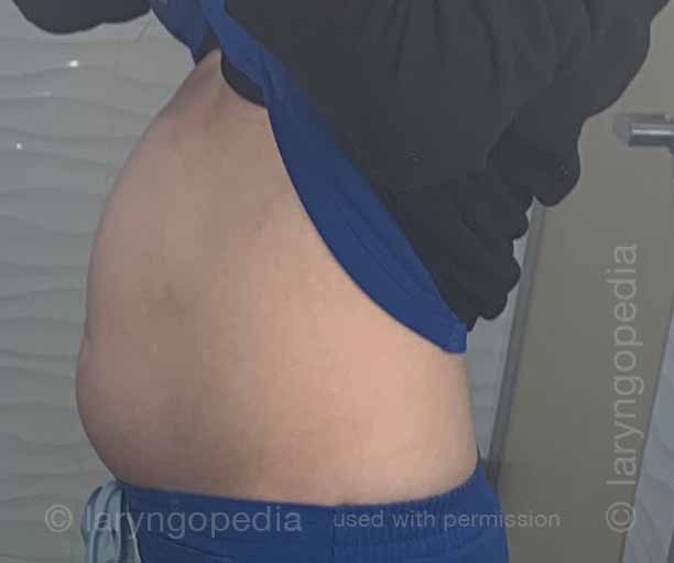
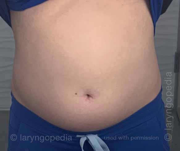
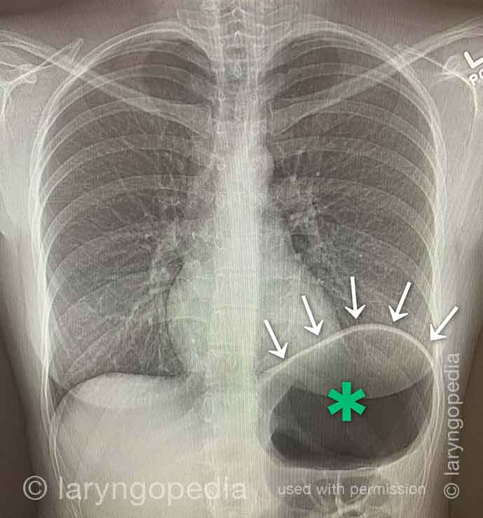
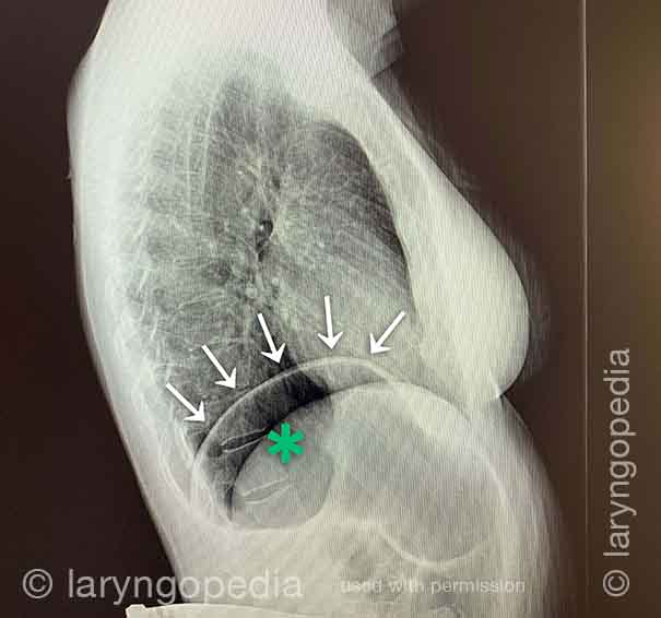
Questa giovane donna ha i classici sintomi R-CPD: la sindrome dell’impossibilità di ruttare. All’inizio della giornata, i suoi sintomi sono minimi e l’addome è al “baseline” perché si è “sgonfiata” a causa della flatulenza durante la notte. In questa serie si vede la differenza nella sua distensione addominale tra l’inizio e la fine della giornata. Le immagini a raggi X mostrano la notevole quantità di aria trattenuta che spiega il suo gonfiore e distensione. La sua progressione è abbastanza tipica; alcuni con R-CPD si distendono anche più di quanto mostrato qui, specialmente dopo aver mangiato un pasto abbondante o consumato qualcosa di gassato.
Side view of a bloated abdomen (1 of 6)
Side view of a bloated abdomen (1 of 6)
Front view (2 of 6)
Front view (2 of 6)
Greater Distention (3 of 6)
Greater Distention (3 of 6)
Front view of bloating stomach (4 of 6)
Front view of bloating stomach (4 of 6)
X-ray of trapped air (5 of 6)
X-ray of trapped air (5 of 6)
Side view (6 of 6)
Side view (6 of 6)
Mancanza di respiro causata da No-Burp (R-CPD)
Le persone che non possono ruttare e hanno la sindrome R-CPD conclamata spesso dicono che quando il gonfiore e la distensione sono particolarmente gravi, e specialmente quando hanno un senso di pressione toracica, hanno anche una sensazione di mancanza di respiro. Diranno, ad esempio, “Sono un [cantante, o corridore, o ciclista o _____], ma le mie capacità sono così ridotte dall’R-CPD. Se gareggio o mi esibisco non posso mangiare o bere per 6 ore prima.” Alcuni dicono addirittura che non possono completare uno sbadiglio quando i sintomi sono particolarmente gravi. I raggi X sottostanti spiegano come l’incapacità di ruttare possa causare mancanza di respiro.
