Incapacidad Para Eructar

La incapacidad para eructar o eructar ocurre cuando el esfínter esofágico superior (músculo cricofaríngeo) no puede relajarse para liberar la «burbuja» de aire. El esfínter es una válvula muscular que rodea el extremo superior del esófago justo debajo del extremo inferior del conducto de la garganta. Si mira de frente el cuello de una persona, está justo debajo de la “nuez de Adán/Eva”, directamente detrás del cartílago cricoides.

Si desea ver esto en un modelo, mire las fotos a continuación. Ese músculo del esfínter se relaja durante aproximadamente un segundo cada vez que tragamos saliva, comida o bebida. Todo el resto del tiempo se contrata. Siempre que una persona eructa, el mismo esfínter necesita soltarse durante una fracción de segundo para que el exceso de aire escape hacia arriba. En otras palabras, así como es necesario que el esfínter se “suelte” para admitir alimentos y bebidas hacia abajo en el acto normal de la deglución, también es necesario que el esfínter pueda “soltar” para dejar salir el aire hacia arriba para eructar. El nombre formal de este trastorno es disfunción cricofaríngea retrógrada (R-CPD).

Las personas que no pueden liberar aire hacia arriba son miserables. Pueden sentir la «burbuja» asentada en la parte media o baja del cuello sin ningún lugar adonde ir. O experimentan gorgoteos cuando el aire sube por el esófago y descubren que la vía de escape está bloqueada por un esfínter que no se relaja. Es como si el músculo del esófago se agitara y apretara continuamente sin éxito.

La persona desea y necesita tanto eructar, pero continúa experimentando esta incapacidad para eructar. A veces esto puede incluso ser doloroso. Estas personas a menudo experimentan presión en el pecho o distensión abdominal, e incluso distensión abdominal. La flatulencia también es grave en la mayoría de las personas con R-CPD. Otros síntomas menos universales son náuseas después de comer, hipo doloroso, hipersalivación o una sensación de dificultad para respirar con el esfuerzo cuando está «lleno de aire». Muchas personas con R-CPD se han sometido a extensas pruebas y tratamientos sin obtener ningún beneficio. R-CPD reduce la calidad de vida y, a menudo, es socialmente perturbador y provoca ansiedad. Los diagnósticos comunes (incorrectos) son «reflujo ácido» y «síndrome del intestino irritable» y, por lo tanto, los tratamientos para estas afecciones no alivian los síntomas de manera significativa.


Enfoques para tratar la incapacidad para eructar:

Para personas que coincidan con el síndrome de:

  1. Incapacidad para eructar
  2. Ruidos de gorgoteo
  3. Presión e hinchazón en el pecho/abdominal
  4. Flatulencia

Esta es la forma más eficiente de avanzar: primero, una consulta para determinar si se cumplen o no los criterios para diagnosticar R-CPD. A continuación, un simple estudio de deglución videoendoscópico en el consultorio que incorpora un examen neurológico de la lengua, la faringe (garganta) y los músculos de la laringe y, a menudo, incluye una miniesofagoscopia. Esto establece que el esfínter funciona normalmente en una dirección de deglución hacia adelante (anterógrada), pero no en una dirección de eructo o regurgitación inversa (retrógrada). Junto con los síntomas descritos anteriormente, esta sencilla consulta en el consultorio y la evaluación de la deglución establecen el diagnóstico de disfunción cricofaríngea retrógrada (falta de relajación).

El segundo paso es colocar Botox en el músculo del esfínter que funciona mal. El efecto deseado del Botox en el músculo es debilitarlo durante al menos varios meses. La persona tiene así muchas semanas para comprobar que el problema se soluciona o al menos se minimiza.

La inyección de Botox podría potencialmente realizarse en un consultorio, pero recomendamos la primera vez (al menos) colocarla durante una breve anestesia general en un quirófano ambulatorio. Eso es porque la primera vez, es importante responder a la pregunta de manera definitiva, es decir, que el problema es la incapacidad del esfínter para relajarse cuando se le presenta una burbuja de aire desde abajo. Además, según una experiencia con más de 1020 pacientes en julio de 2022, una sola inyección parece «entrenar» al paciente a eructar. Aproximadamente el 80% de los pacientes han mantenido la capacidad de eructar mucho después de que se haya disipado el efecto del Botox. Es decir, mucho más allá de los 6 meses desde el momento de la inyección.

Los pacientes tratados por R-CPD como se acaba de describir deberían experimentar un alivio espectacular de sus síntomas. Y para repetir, nuestra experiencia en el tratamiento de más de 1020 pacientes (y contando) sugiere que esta sola inyección de Botox permite que el sistema se «reinicie» y es posible que la persona nunca pierda su capacidad para eructar. Por supuesto, si el problema reaparece, la persona podría optar por seguir tratamientos adicionales con Botox, o incluso podría optar por someterse a una miotomía cricofaríngea con láser endoscópica. Para obtener más información sobre esta afección, consulte la descripción del Dr. Bastian de su experiencia con los primeros 51 de su número de casos mucho mayor.

Seminario web R-CPD + preguntas y respuestas

complications of botox injections for R-CPD under general anesthesia YT Thumbnail
Reproducir vídeo
Teleconversación
con el Dr. Bastian
¿Todavía tienes preguntas?
Programe una llamada con el
Dr. Bastian a través de Zoom.

Lifting the CPM for a R-CPD Injection

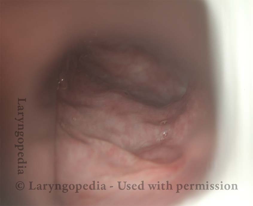
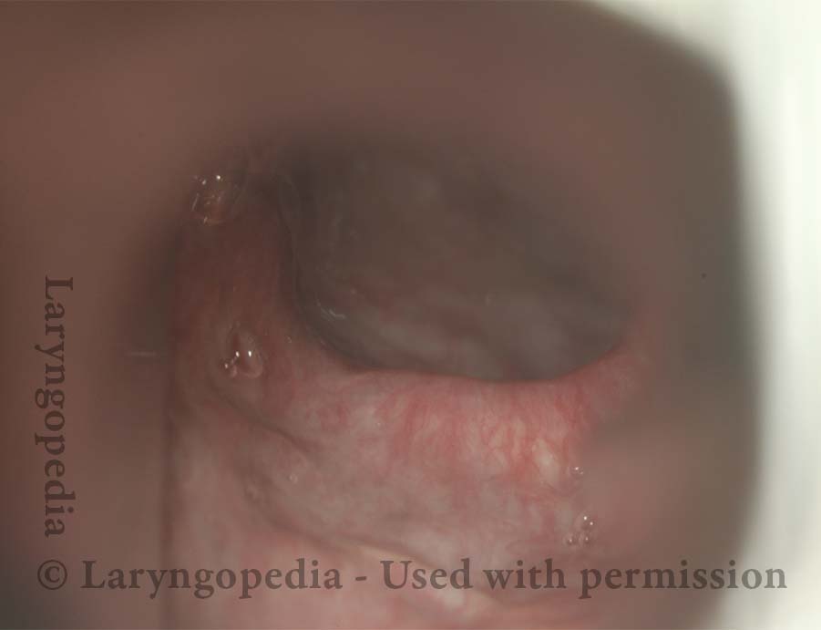
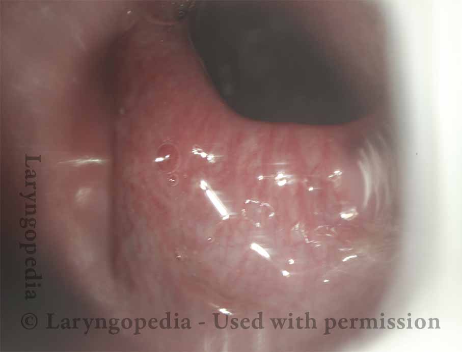
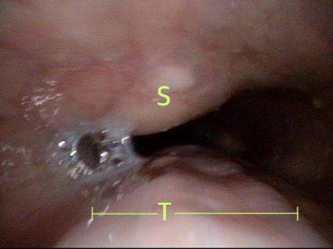
These are intra-operative photos of one of nearly 1500 persons treated for R-CPD as of September 2023. This sequence shows several things:   The dilated, “always open” esophagus distal (below) the muscle; how to identify the cricopharyngeus muscle; and one way of injecting it.

Visual Portfolio, Posts & Image Gallery para WordPress

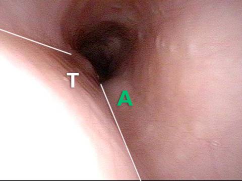
Above the CPM (1 of 5)

The view here is just inside the uppermost part of the esophagus, immediately behind the larynx. The target muscle is not yet seen, but the dilation of the esophagus is notable in this young, muscular male.

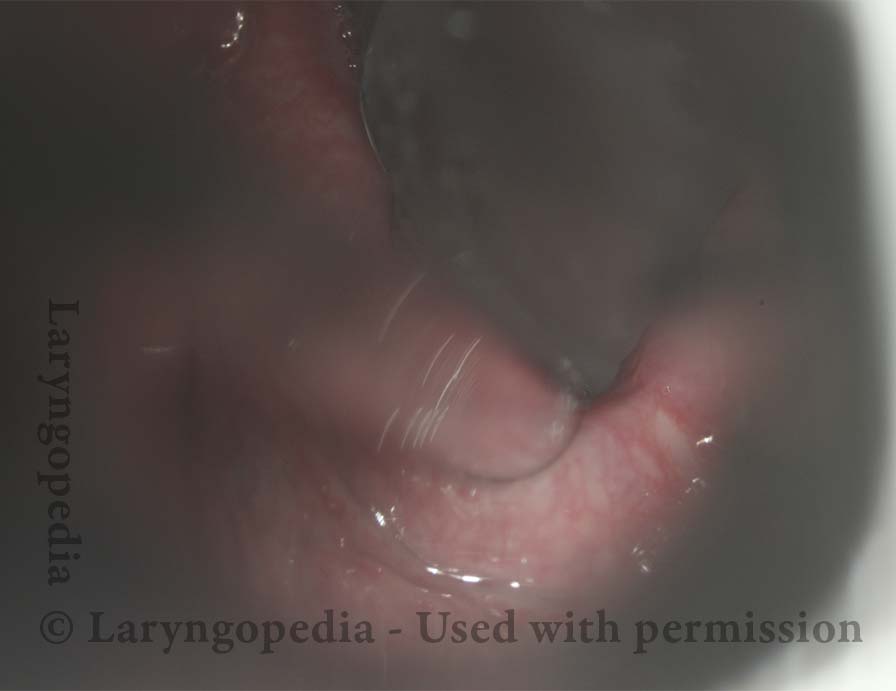
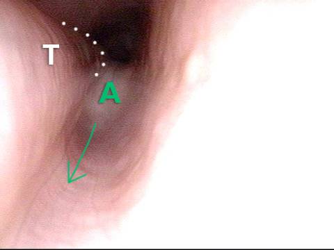
Ridge of the CPM (2 of 5)

The larynx is gently lifted forward, and the “ridge” of the muscle begins to be seen.

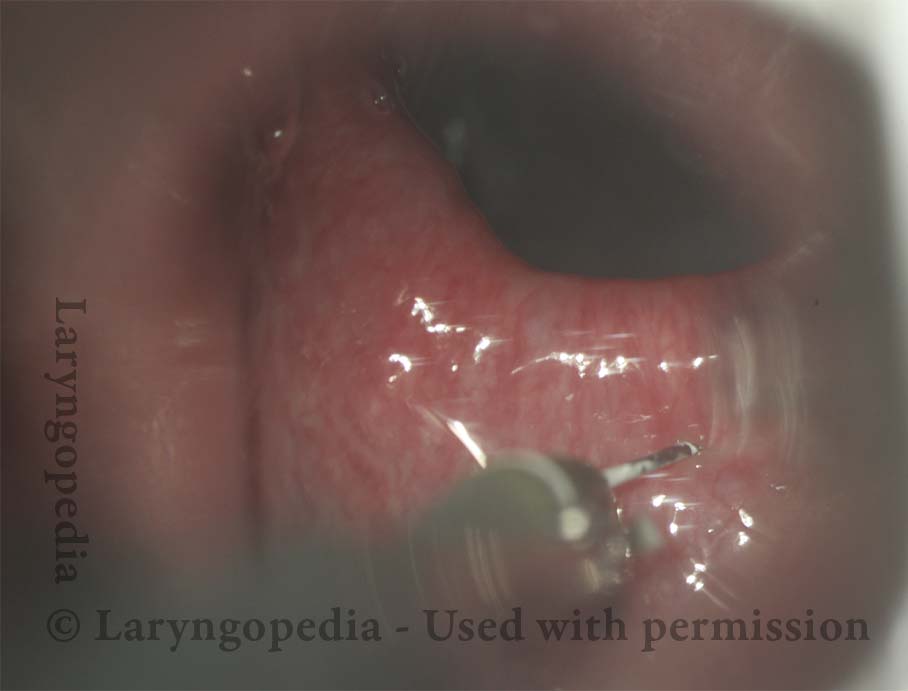
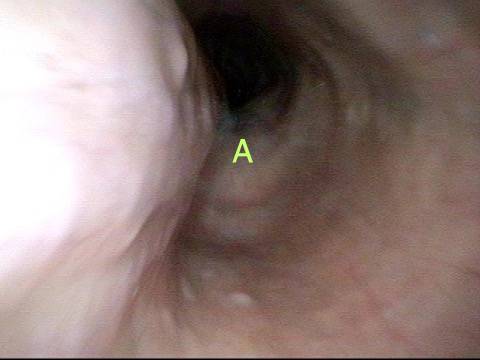
Exposed CPM ( 3 of 5)

With further lifting, the muscle becomes obvious and is ready for injection.

CPM Palpated ( 4 of 5)

If desired, the muscle can be palpated—here with the side of a suction cannula—to determine compressibility and “size.”

Botox injection ( 5 of 5)

A 25-gauge butterfly has had one “wing” cut off completely, and the other wing cut to leave only a nubbin for grasping with a straight alligator forceps. Seen here is injection of one of three or four sites, chosen to distribute the botox throughout the entire muscle.

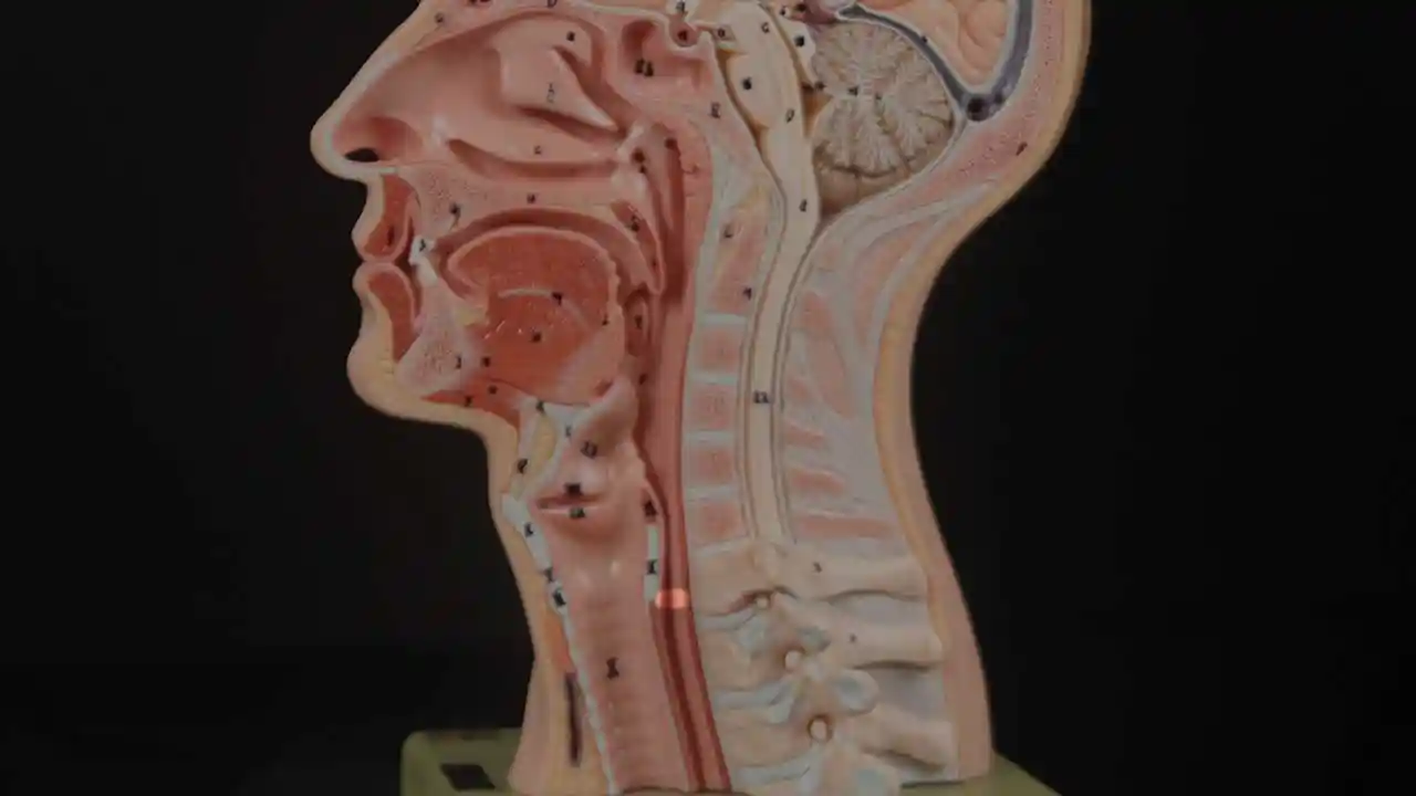
¿Dónde está el músculo cricofaríngeo?

Visual Portfolio, Posts & Image Gallery para WordPress

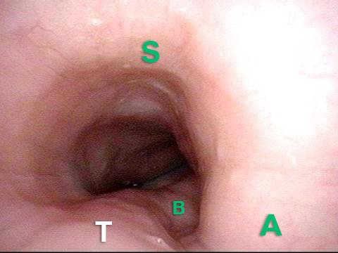
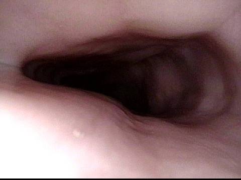
Cricopharyngeus Muscle (1 of 3)

The highlighted oval represents the location of the cricopharyngeus muscle.

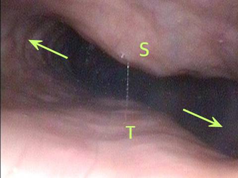
Open Cricopharyngeus Muscle (2 of 3)
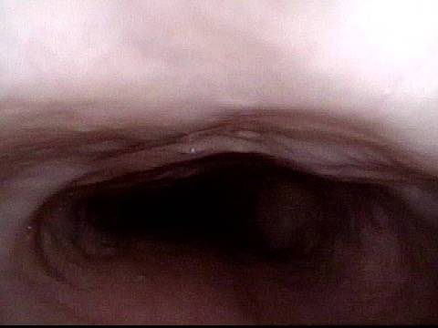
The cricopharyngeus muscle in the open position.

Closed (3 of 3)

The cricopharyngeus muscle in the contracted position.

Hallazgos Esofágicos

Visual Portfolio, Posts & Image Gallery para WordPress

Aortic shelf (1 of 3)

A view of the mid-esophagus in a young person (early 30’s). The esophagus is kept open by the patient’s un-burped air. Note the “aortic shelf” at A, delineated by dotted lines.

Bony spur emerges due to stretched esophagus (2 of 3)

A moment later, additional air is pushed upwards from the stomach to dilate the mid-esophagus even more. A bony “spur” in the spine is thrown into high relief by the stretched esophagus.

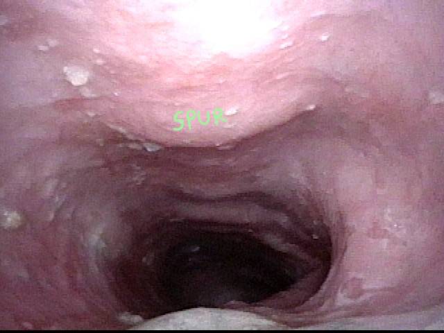
Stretched esophagus due to unburpable air (3 of 3)

A view of the upper esophagus (from just below the cricopharyngeus muscle sphincter) shows what appears to be remarkable lateral dilation (arrows) caused over time by the patient’s unburpable air. Dilation can only occur laterally due to confinement of the esophagus by trachea (anteriorly) and spine (posteriorly), as marked.

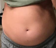
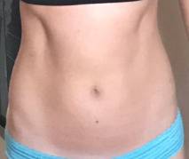
Distensión Abdominal de R-CPD

Visual Portfolio, Posts & Image Gallery para WordPress

Gastric Air Bubble (1 of 3)

This abdominal xray of an individual with R-CPD shows a remarkably large gastric air bubble (dotted line), and also excessive air in transverse (T) and descending (D) colon. All of this extra air can cause abdominal distention that increases as the day progresses.

Bloated Abdomen (2 of 3)

Flatulence in the evening and even into the night returns the abdomen to normal, but the cycle repeats the next day. To ask patients their degree of abdominal distention, we use pregnancy as an analogy in both men and women. Not everyone describes this problem. Most, however, say that late in the day they appear to be “at least 3 months pregnant.” Some say “6 months” or even “full term.” In a different patient with untreated R-CPD, here is what her abdomen looked like late in every day. Her abdomen bulges due to all of the air in her GI tract, just as shown in Photo 1.

Non-bloated Abdomen (3 of 3)

The same patient, a few weeks after Botox injection. She is now able to burp. Bloating and flatulence are remarkably diminished, and her abdomen no longer balloons towards the end of every day.

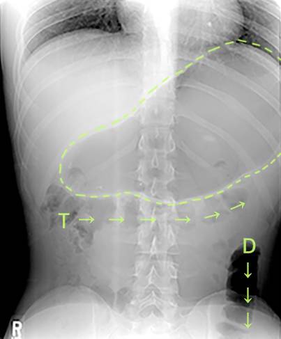
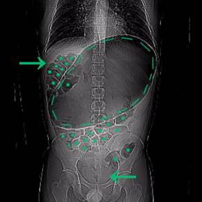
Una Rara «crisis abdominal» Debido a R-CPD (incapacidad para eructar)

R-CPD in X-ray Pictures: Misery vs. Crisis from Inability to Burp
Reproducir video acerca de R-CPD in X-ray Pictures: Misery vs. Crisis from Inability to Burp

Este joven tenía una crisis abdominal relacionada con R-CPD. Ha tenido síntomas de por vida de R-CPD clásico: incapacidad para eructar, gorgoteo, hinchazón y flatulencia. Durante un momento de particular incomodidad, lamentablemente tomó un “remedio” que era carbonatado. Aquí se ve una enorme burbuja de aire en el estómago. Muchos de sus intestinos están llenos de aire y presionados hacia arriba y hacia su derecha (a la izquierda de la foto, en la flecha). La presión interna dentro de su abdomen también cortó su capacidad de expulsar gases.

Visual Portfolio, Posts & Image Gallery para WordPress

X-Ray of Abdominal Bloating (1 of 2)

Note arrow pointing to lack of gas in the descending colon/rectum. NG decompression of his stomach allowed him to resume passing gas, returning him to his baseline “daily misery” of R-CPD.

Original X-Ray (2 of 2)

X-Ray without markings

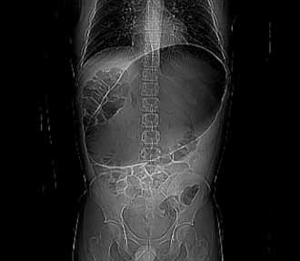
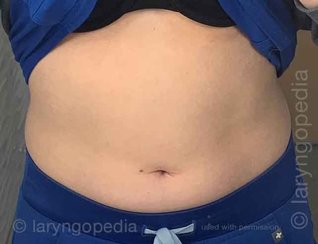
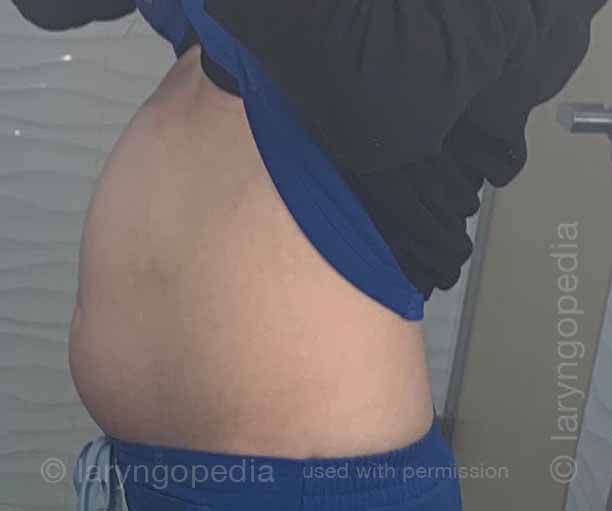
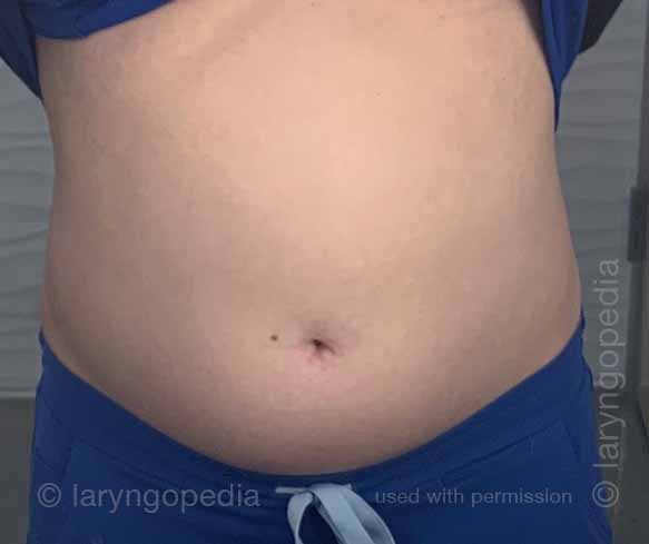
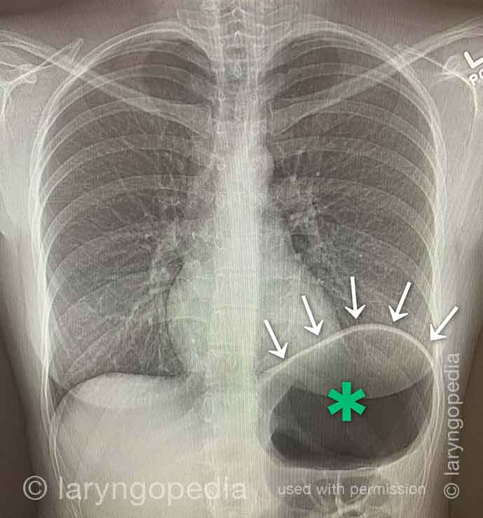
No puedo eructar: Progresión de la hinchazón y la distensión abdominal; Un ciclo diario para muchos con R-CPD

Esta joven tiene los síntomas clásicos de R-CPD: el síndrome de no poder eructar. Temprano en el día, sus síntomas son mínimos y el abdomen está en la «línea de base» porque se ha «desinflado» a través de la flatulencia durante la noche. En esta serie se ve la diferencia en su distensión abdominal entre temprano y tarde en el día. Las imágenes de rayos X muestran la notable cantidad de aire retenido que explica su hinchazón y distensión. Su progresión es bastante típica; algunos con R-CPD se distienden incluso más de lo que se muestra aquí, especialmente después de comer una comida copiosa o consumir algo carbonatado.

Visual Portfolio, Posts & Image Gallery para WordPress

Side view of a bloated abdomen (1 of 6)

Early in the day, side view of the abdomen shows mild distention. The patient’s discomfort is minimal at this time of day as compared with later.

Front view (2 of 6)

Also early in the day, a front view, showing again mild distention.

Greater Distention (3 of 6)

Late in the same day, another side view to compare with photo 1. Accumulation of air in stomach and intestines is distending the abdominal wall.

Front view of bloating stomach (4 of 6)

Also late in the day, the front view to compare with photo 2, showing considerably more distention. The patient is quite uncomfortable, bloated, and feels ready to “pop.” Flatulence becomes more intense this time of day, and will continue through the night.

X-ray of trapped air (5 of 6)

Antero-posterior xray of the chest shows a very large stomach air bubble (at *) and the descending colon is filled with air (arrow).

Side view (6 of 6)

A lateral view chest xray shows again the large amount of excess air in the stomach and intestines that the patient must rid herself of via flatulence, typically including through the night, in order to begin the cycle again the next day.

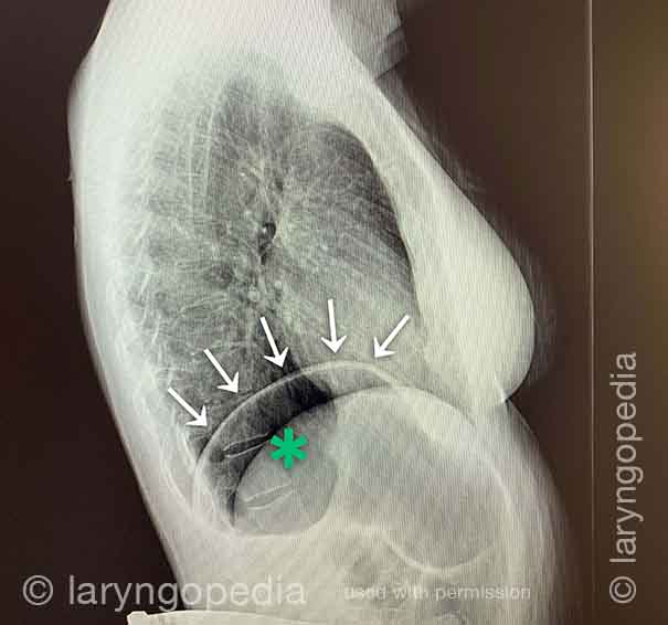
Dificultad para Respirar Causada por no Eructar (R-CPD)

Las personas que no pueden eructar y tienen el síndrome R-CPD completo a menudo dicen que cuando la hinchazón y la distensión son particularmente malas, y especialmente cuando tienen una sensación de presión en el pecho, también tienen una sensación de dificultad para respirar. Dirán, por ejemplo, “Soy un [cantante, o corredor, o ciclista o _____], pero mi habilidad está muy disminuida por R-CPD. Si estoy compitiendo o actuando, no puedo comer ni beber durante las 6 horas anteriores”. Algunos incluso dicen que no pueden completar un bostezo cuando los síntomas son particularmente graves. Las radiografías a continuación explican cómo la incapacidad para eructar puede causar dificultad para respirar. 

Visual Portfolio, Posts & Image Gallery para WordPress

X-ray of trapped air (1 of 2)

In this antero-posterior xray, one can see that there is so much air in the abdomen, that the diaphragm especially on the left (right of xray) is lifted up, effectively diminishing the volume of the chest cavity and with it, the size of a breath a person can take.

Side view (2 of 2)

The lateral view again shows the line of the thin diaphragmatic muscle above the enormous amount of air in the stomach. The diaphragm inserts on itself so that when it contracts it flattens. That action sucks air into the lungs and simultaneously pushes abdominal contents downward. But how can the diaphragm press down all the extra air? It can’t fully, and the inspiratory volume is thereby diminished. The person says “I can’t get a deep breath.”

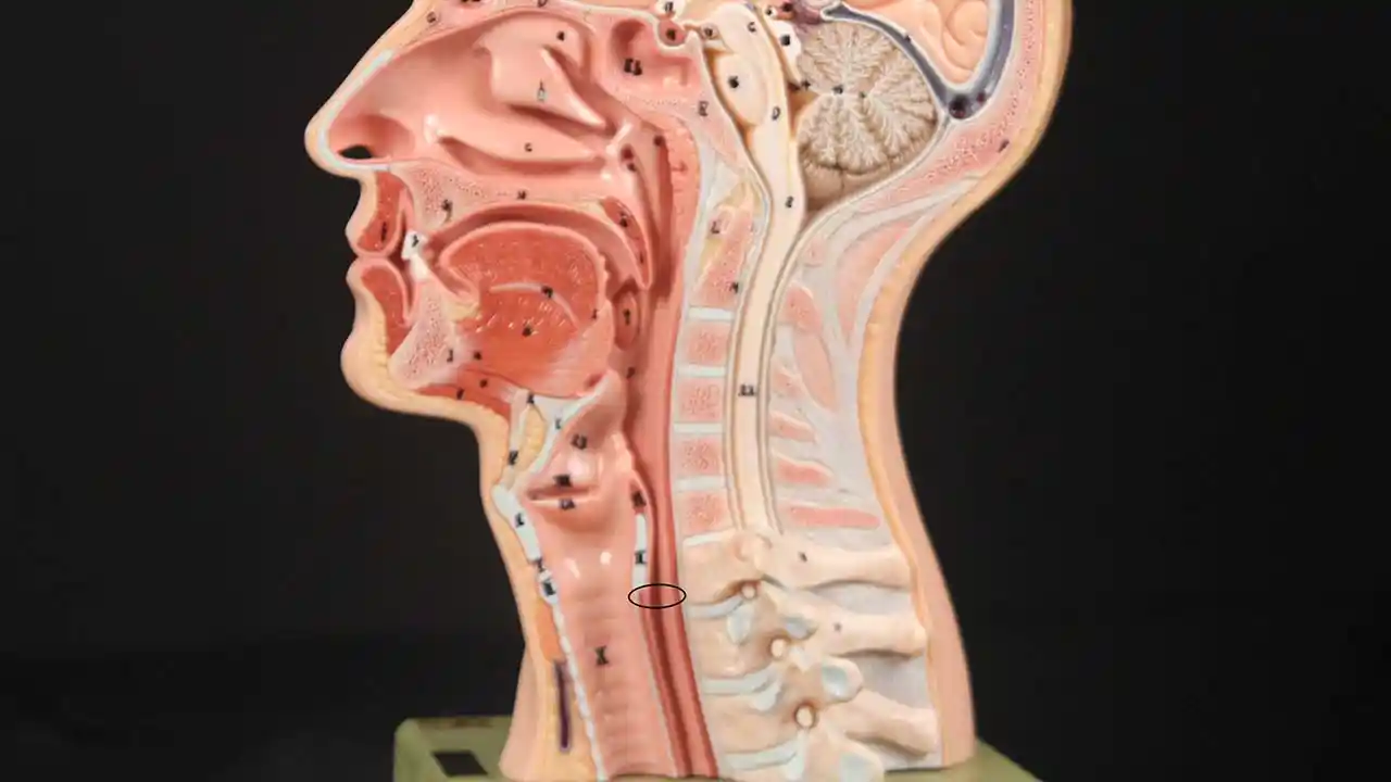
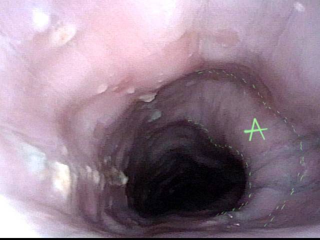
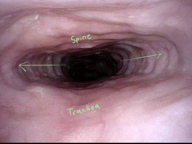
Hallazgos Esofágicos más Interesantes de R-CPD (incapacidad para eructar)

Visual Portfolio, Posts & Image Gallery para WordPress

Stretched Esophagus (1 of 4)

Using a 3.7mm ENT scope with no insufflated air, note the marked dilation of the esophagus by swallowed air the patient is unable to belch. T = trachea; A = aortic shelf; S = spine

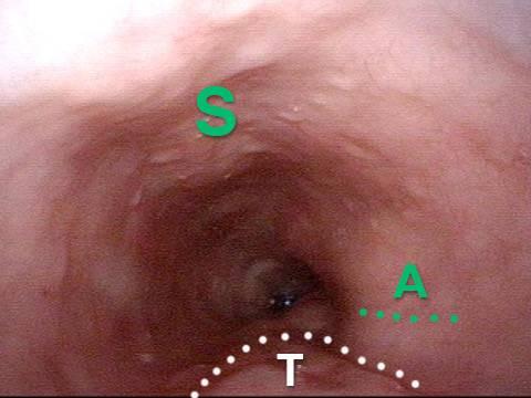
Tracheal Wall (2 of 4)

The posterior wall of the trachea (T) is better seen here from a little higher in the esophagus. A = aorta

Over-dilation (3 of 4)

The photo is rotated clockwise at a moment when air from below is pushed upward so as to transiently over-dilate the esophagus. Note that the esophagus is almost stretching around the left side of the trachea in the direction of the arrow.

Bronchus (4 of 4)

Now deeper in the esophagus (with it inflated throughout the entire examination by the patient’s own air), it even appears that the left mainstem bronchus (B) is made visible by esophageal dilation stretching around it.

Dramática Dilatación Lateral del Esófago Superior

Visual Portfolio, Posts & Image Gallery para WordPress

Remarkable lateral dilation (1 of 3)

This photo is at the level of (estimated) C6 of the spine (at S). This person has known cervical arthritis, accounting for the prominence. Opposite the spine is the trachea (T). Note the remarkable lateral dilation (arrows) in this picture obtained with with no insufflated air using a 3.6mm ENF-VQ scope. It is the patient’s own air keeping the esophagus open for viewing.

Dilated upper esophagus (2 of 3)

At a moment when air from below further dilates the upper esophagus, the tracheal outline is particularly well-seen (T) opposite the spine (S). The “width” of the trachea indicated further emphasizes the degree of lateral dilation, which is necessary because spine and trachea resist anteroposterior dilation.

Aortic shelf (3 of 3)

Just for interest, at mid-esophagus, the familiar aortic “shelf” is seen. Again, this esophagus is being viewed with a 3.6 mm scope with only the patient own (un-burped) air allowing this view.

Cómo Puede Verse el Esófago «debajo de un eructo”

Visual Portfolio, Posts & Image Gallery para WordPress

Baseline (1 of 3)

Mid-esophagus of a person with R-CPD who is now burping well after Botox injection into the cricopharyngeus muscle many months earlier. The esophagus remains somewhat open likely due to esophageal stretching from the years of being unable to burp and also a “coming burp.”

Pre-burp (2 of 3)

A split-second before a successful burp the esophagus dilates abruptly from baseline (photo 1) as the excess air briefly enlarges the esophagus. An audible burp occurs at this point.

Post-burp (3 of 3)

The burp having just happened, the esophagus collapses to partially closed as the air that was “inflating it” has been released.

¿Desde Dónde han Viajado los no-burpers?

Subscribe
Notify of

6 Comments
Newest
Oldest Most Voted
Inline Feedbacks
View all comments
6
0
Click to see all comments.x