An encyclopedia about voice, swallowing, airway, coughing, & other head + neck disorders.

Polipi delle Corde Vocali

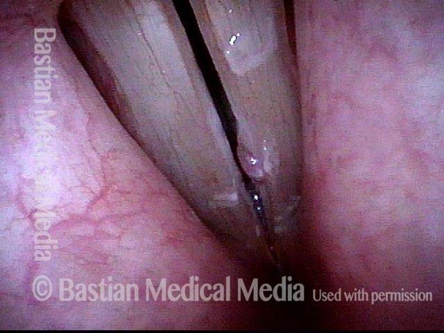
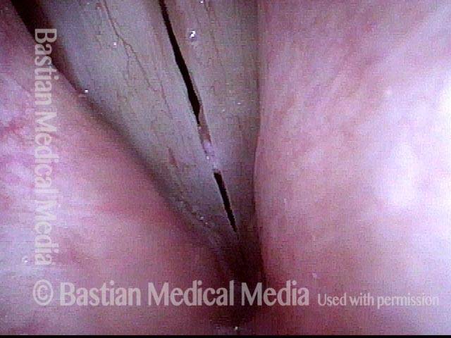
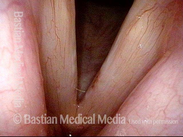
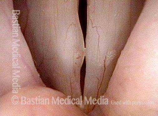
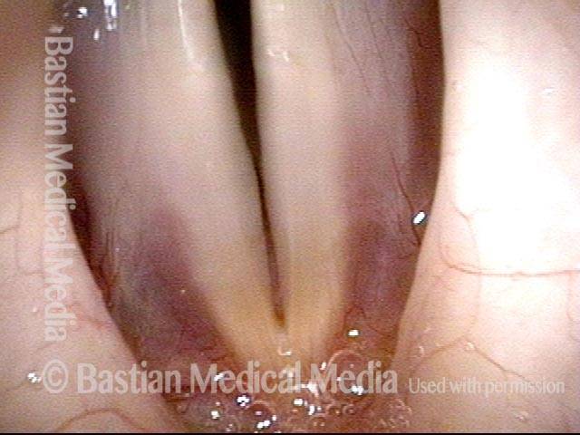
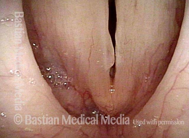
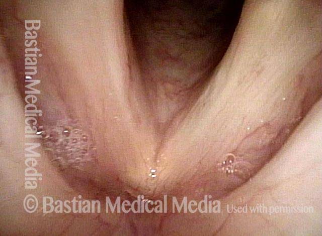
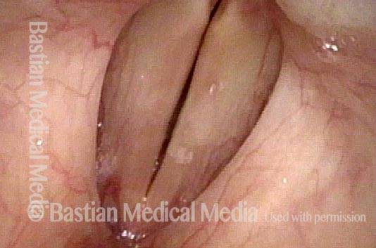
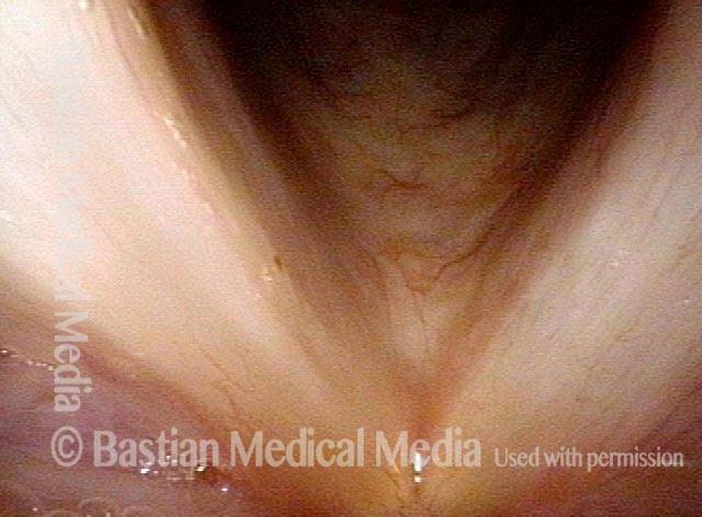
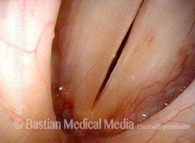
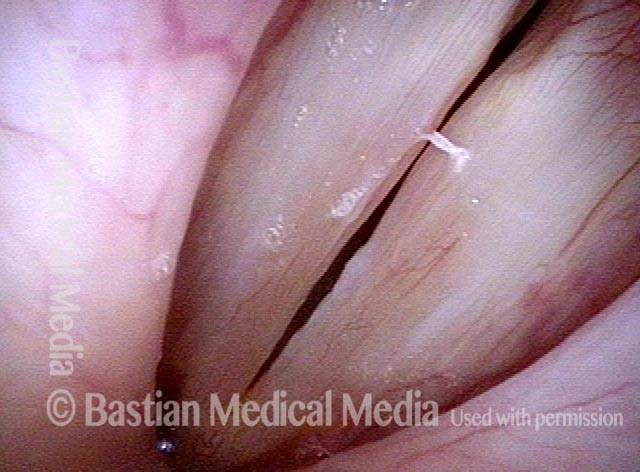
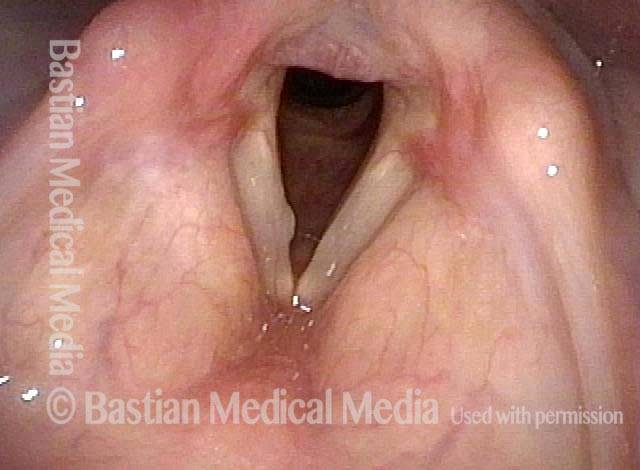
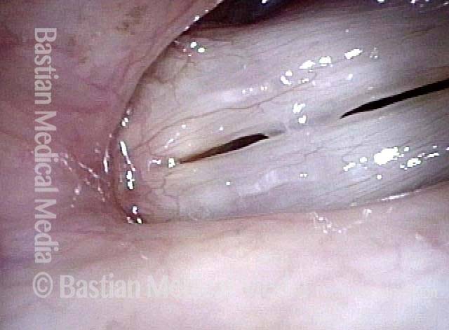
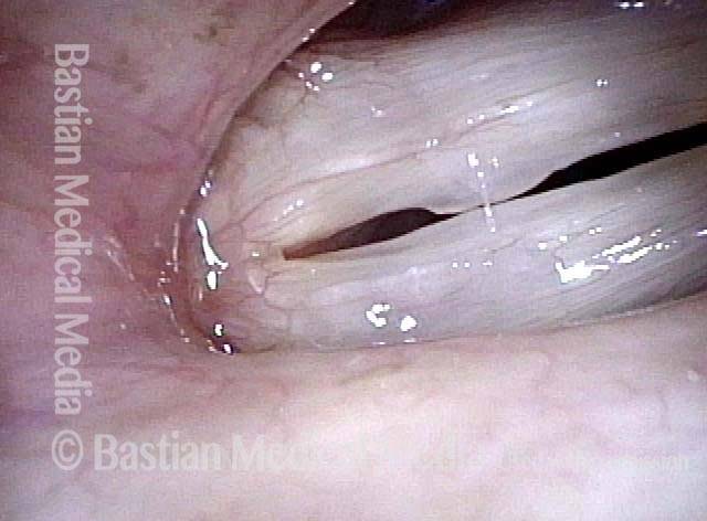
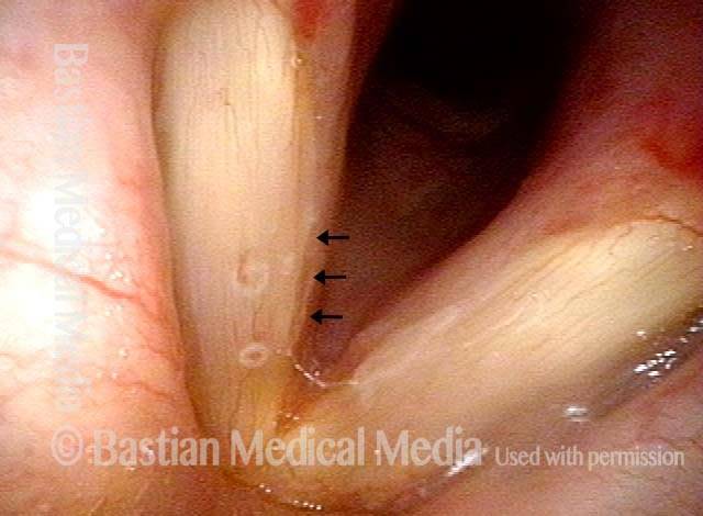
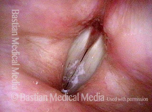
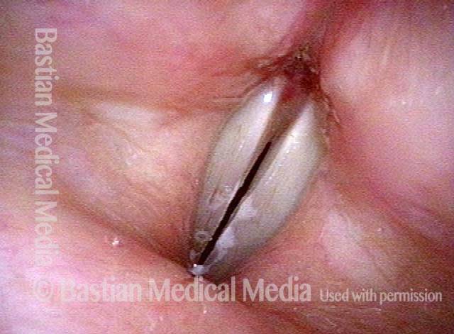
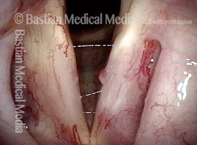
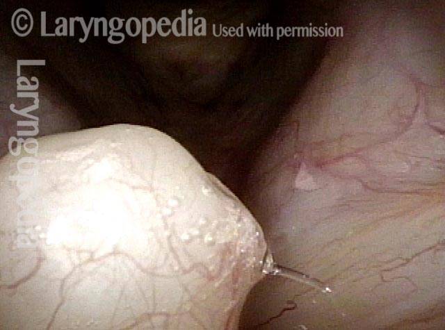
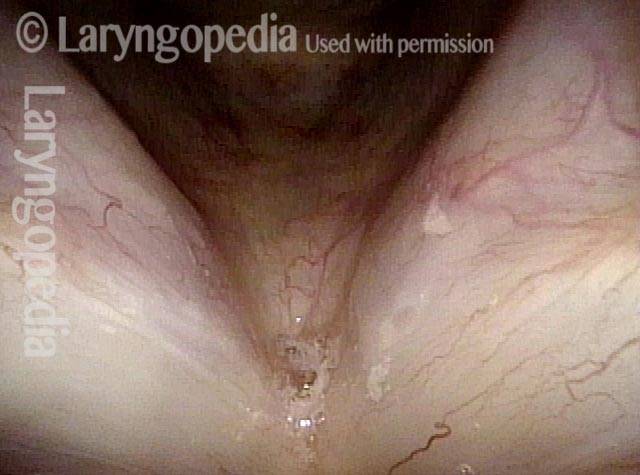
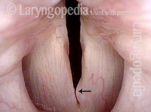
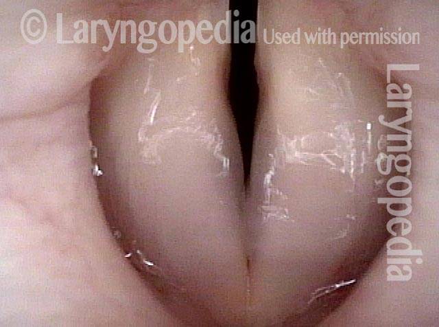
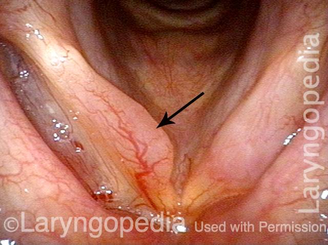
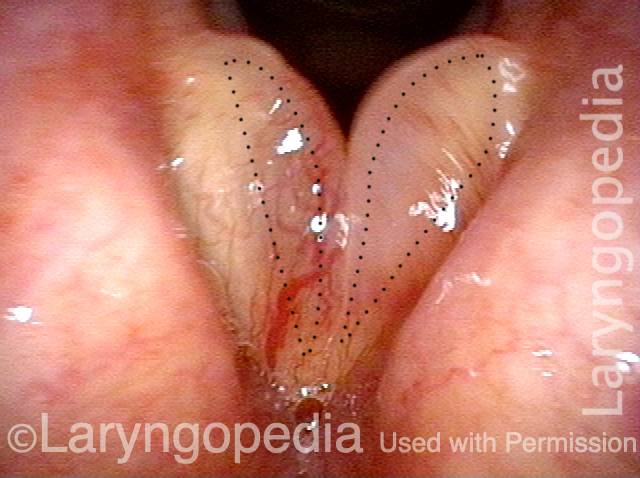
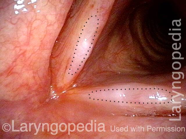
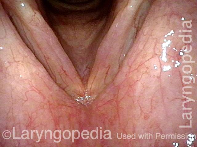
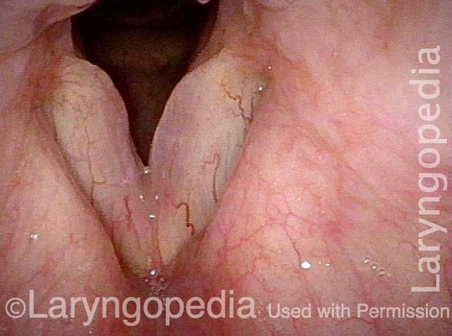
I polipi sono un grande rigonfiamento sulla corda vocale che tipicamente si verifica unilateralmente, cioè senza un rigonfiamento simile sulla corda opposta. Il termine polipo vocale è alquanto impreciso, ma i polipi vocali possono essere distinti da un tipo simile di gonfiore, i noduli vocali, in almeno due modi:

  1. I polipi tendono ad essere più grandi dei noduli.
  2. I polipi si presentano unilateralmente o sono notevolmente più grandi di una lesione della corda vocale opposta, mentre i noduli si presentano in coppie e sono generalmente di dimensioni simili.

I polipi e i noduli vocali sono causati almeno in parte da traumi vibratori, dovuti a un uso eccessivo della voce acuto (con polipi) o cronico.

Un polipo vocale disturba la chiarezza della voce e altre capacità interferendo con l’approssimazione accurata delle corde vocali durante la fonazione. Un polipo può anche aggiungere massa alle corde vocali, riducendo così la gamma tonale disponibile per la voce. I polipi possono essere definiti emorragici, peduncolati e così via.


Polipi vocali rimossi e poi ricorrenti

Visual Portfolio, Posts & Image Gallery for WordPress

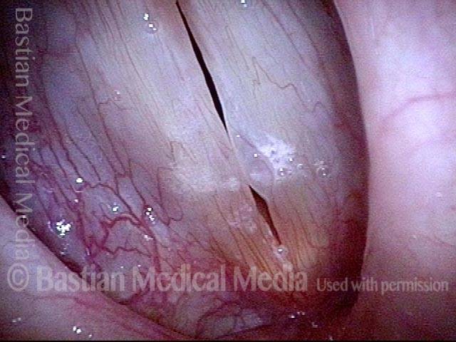
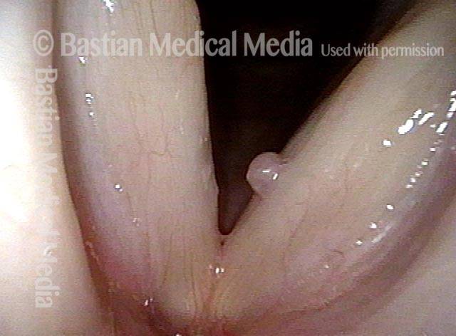
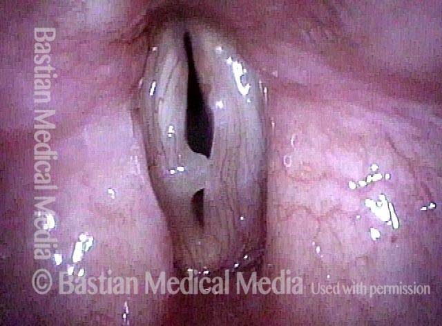
Vocal polyp (1 of 4)

The translucency of the left vocal cord polyp (right of image) and the patient’s long history of hoarseness together suggest chronicity. Initial voice therapy to manage the patient’s obvious “vocal overdoer” status.

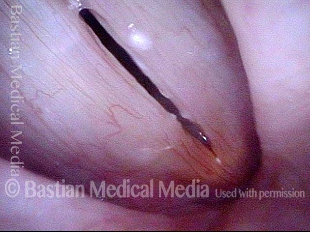
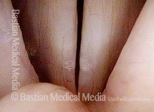
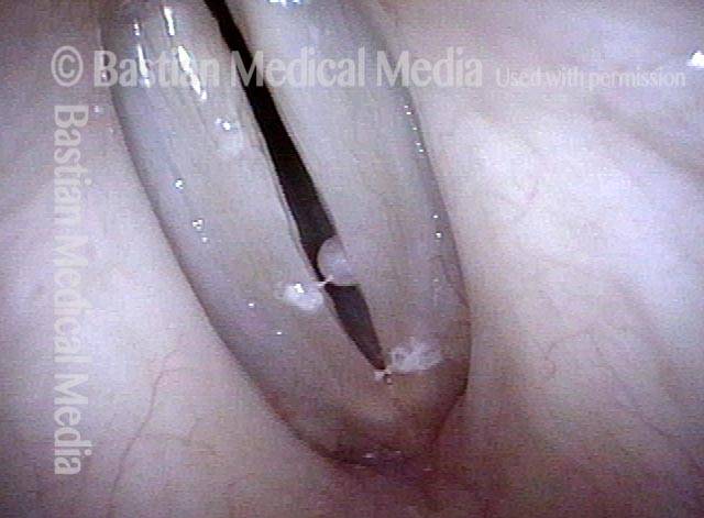
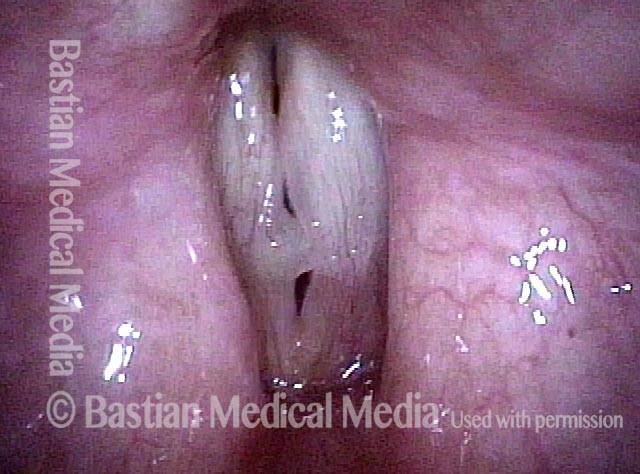
Vocal polyp (2 of 4)

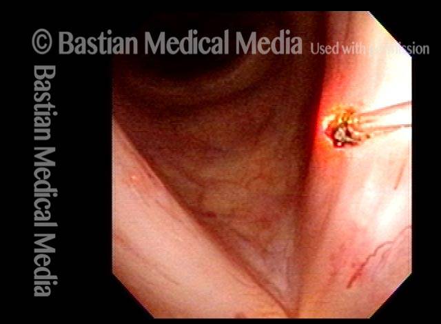
Additional view, at diagnosis.

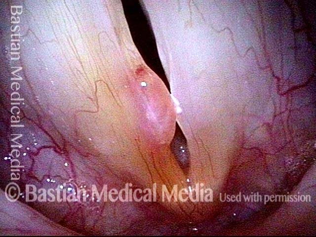
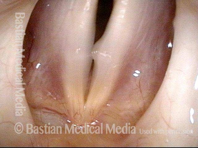
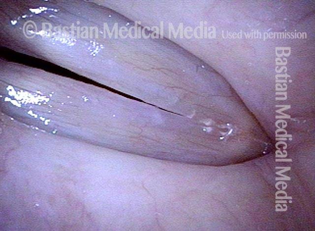
Vocal polyp, one week after surgical removal (3 of 4)

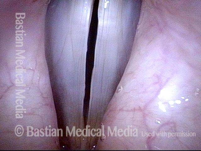
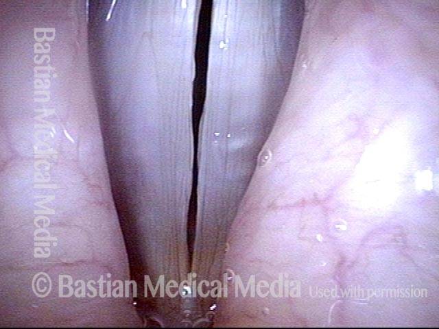
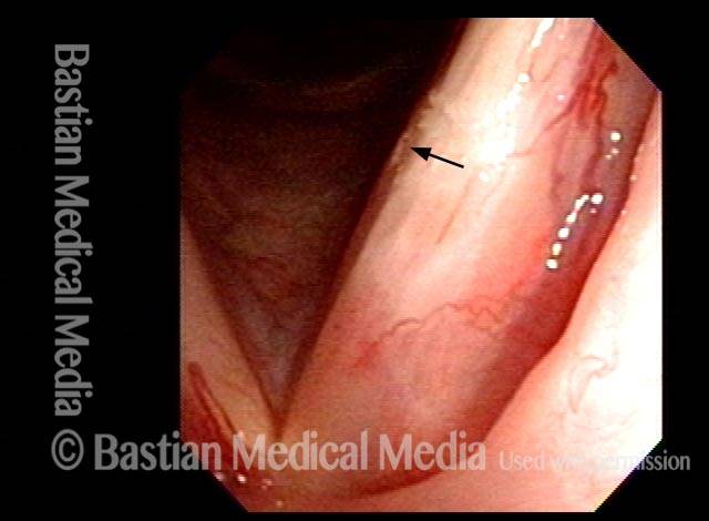
Representative view, one week after microsurgical removal of left vocal cord polyp (right of image). Strobe light, open phase of vibration, with bilaterally equal free margin and phonatory “match.”

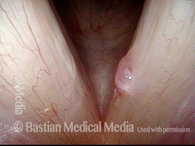
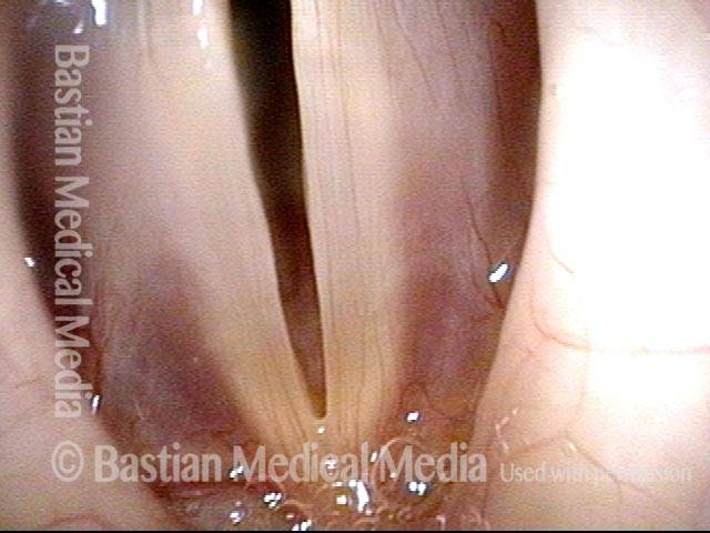
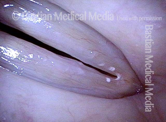
Vocal polyp, subsequent new injury (4 of 4)

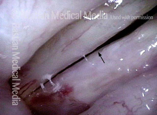
Approximately two months later, new injury from persistent vocal overuse.

Polipi vocali, prima e dopo l’intervento chirurgico

Visual Portfolio, Posts & Image Gallery for WordPress

Vocal polyp (1 of 6)

An operatic baritone has a chronic left vocal cord polyp (right of image), and small contact reaction, right cord.

Vocal polyp (2 of 6)

Phonation, open phase of vibration, upper middle voice, showing obvious margin elevation of the left cord (right of image). Voice is hoarse.

Vocal polyp (3 of 6)

Maximum closed phase of vibration, showing polyp-induced gap, causing air wasting and hoarse voice quality.

Vocal polyp, surgically removed (4 of 6)

Sixth day after microlaryngoscopic removal of the polyp. Note the red, 2-millimeter “wound” where the polyp was removed.

Vocal polyp, surgically removed (5 of 6)

At extremely high falsetto, open phase of vibration, showing uniform width of glottic chink. Voice is normal, even at this revealing, high pitch.

Vocal polyp, surgically removed (6 of 6)

Closed phase of vibration, high falsetto, shows equal vibratory amplitude on both sides (no stiffness) and excellent match of the cords.

Esempio 2

Visual Portfolio, Posts & Image Gallery for WordPress

Vocal polyp (1 of 2)

Chronic polyp on the right vocal cord (left of image), with ectatic capillaries, unresponsive to voice rest and therapy.

Vocal polyp, surgically removed (2 of 2)

Seven weeks after surgical removal and spot coagulation of ectatic capillaries. The margins of the vocal cords now match, and capillaries are normalized. Mucosal vibration is preserved to the highest reaches of the singing range. At this pre-phonatory instant, one can see that muscle memory is keeping the vocal cords slightly apart, suggesting the need for additional speech therapy.

Esempio 3

Visual Portfolio, Posts & Image Gallery for WordPress

Vocal polyp (1 of 6)

Prephonatory instant, standard light. The space between the vocal cords is larger than necessary to accommodate the polyp (right of image) and low-profile elevation (left of image).

Vocal polyp (2 of 6)

Phonation with blurring, standard light.

Vocal polyp, surgically removed (3 of 6)

Six days after surgical removal. Prephonatory instant, standard light. Compare with photo 1. The patient continues to position vocal cords in a surprisingly separated position, as though the ghosts of the swellings remain. We call this “gap memory” or “posture memory,” though it is a manifestation as well of muscular tension dysphonia.

Vocal polyp, surgically removed (4 of 6)

Phonation with blurring, standard light. Compare with photo 2.

Vocal polyp, surgically removed (5 of 6)

Phonation, strobe light, open phase of vibration, at high G# (~831 Hz), just below A5. Even at this high pitch, both cords oscillate out to a full lateral excursion.

Vocal polyp, surgically removed (6 of 6)

Phonation, strobe light, closed phase of vibration, also at high G#. Since the patient is only six days postop, mild residual swelling is still present. More importantly, note that this “closed” vibration phase is not in fact fully closed, as further evidence of the patient’s “gap memory” and muscular tension dysphonia. Singing voice-qualified speech therapy and work with a singing teacher will address this.

Polipo traslucido

Visual Portfolio, Posts & Image Gallery for WordPress

Translucent polyp (1 of 4)

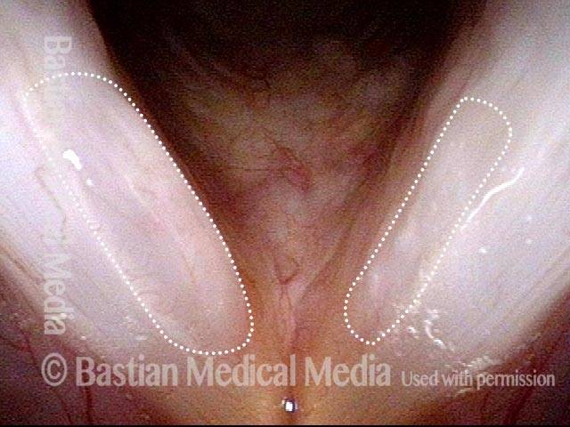
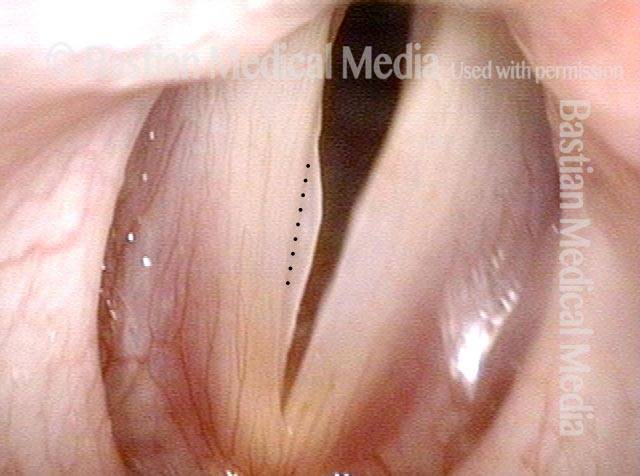
Close-range view with vocal cords in abducted position. This is not the best view to see translucence but faintly “grey” tone of polyps (circled by dotted lines) is indicator of translucence.

Translucent polyp (2 of 4)

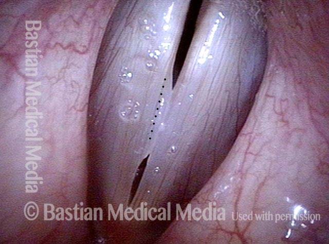
As vocal cords are coming towards adduction, grey indicator of translucence.

Translucent polyp (3 of 4)

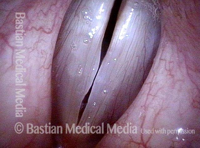
Similar view, with elicitation of rapid inspiration to reveal polyps better, especially on left (right of image).

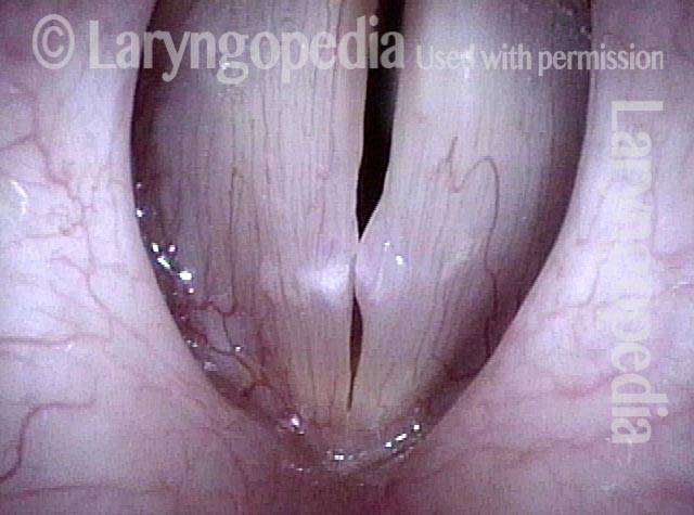
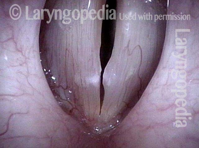
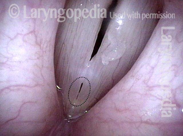
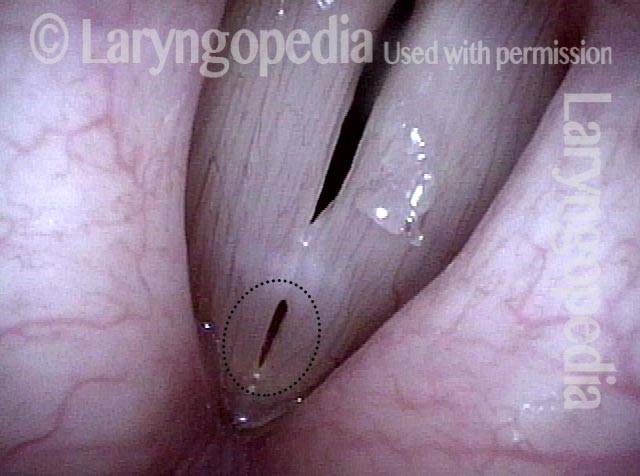
Translucent polyp (4 of 4)

During strobe illumination, translucence especially of the right vocal cord (left of image), is seen best. Note that the larger polyp rides on the margin of the left vocal cord (right of image).

Rimosso polipo di cantante lirico con ripristino delle capacità originarie

Visual Portfolio, Posts & Image Gallery for WordPress

Polyp and capillary ectasia (1 of 8)

Right vocal cord polyp (left of photo) and bilateral capillary ectasia. This young man continues to perform successfully but with increased effort, reduced endurance, impaired falsetto.

Prephonatory instant (2 of 8)

Prephonatory instant (standard light, just before vibratory blur) at high pitch to make the polyp maximally evident.

One week post-op (3 of 8)

One week after microsurgical removal and spot-coagulation of ectasia.

Prephonatory instant (4 of 8)

Prephonatory instant at B4 (494 Hz) (compare with photo 2).

One month post-op (5 of 8)

One month after surgery, original vocal capabilities are restored. A tiny ectatic capillary “escaped.” It can be observed or spot-coagulated in an office setting using the pulsed-KTP laser.

Prephonatory instant (6 of 8)

Prephonatory instant shows straight margins and excellent “margin match.”

Closed phase (7 of 8)

Strobe light, closed phase of vibration. This is performed at E-flat 5 (high falsetto), in order to maximally “reveal” (if present) vibratory stiffness.

Open phase (8 of 8)

Open phase shows equal lateral excursion of both cords: there is no stiffness, and the residual irregularity is on the left (un-operated) cord (right of photo).

Polipo di un’attrice prima e ore dopo la rimozione chirurgica

Visual Portfolio, Posts & Image Gallery for WordPress

Vocal cord polyp (1 of 8)

Musical theater actress with chronic hoarseness due to this right vocal cord polyp (left of photo), first identified a year earlier and unresponsive to speech therapy.

Closer view (2 of 8)

Magnified view shows small elevation of the left cord (right of photo) as well.

Closed phase (3 of 8)

Closed phase of vibration (strobe light) at B flat 5 (932 Hz).

Open phase (4 of 8)

Open phase at same pitch.

24 hours post surgery (5 of 8)

The next day, only a few hours after surgical removal. Note slight bruising from the endotracheal tube (long arrows), and small dots where a laser impact was used to interrupt flow in a prominent capillary (short arrows).

Primary “wound” (6 of 8)

Magnified view. Primary “wound” from polyp removal is at the arrows. Edema of the opposite side is from minimal trimming on that side.

Closed phase (7 of 8)

Closed phase of vibration, strobe light, at D5 (587 Hz). Increased mucus is from endotracheal tube and surgical manipulation just hours earlier.

Open phase (8 of 8)

Open phase, at same pitch. Subtle irregularities will “iron out” within over time. Voice is already much better (tested briefly because the patient is within the four days of voice rest, and then will gradually increase amounts of voice use for the subsequent month).

Il cavo operato ha un aspetto migliore rispetto al cavo non operato

Visual Portfolio, Posts & Image Gallery for WordPress

Singer with chronic hoarseness (1 of 4)

Chronic hoarseness in a singer. The obvious abnormality is on the left vocal cord (right of photo).

Attempting phonation (2 of 4)

When trying to make sound at high pitch, the polyp comes into early contact and interferes with vibration. Chaotic vibration will not allow full evaluation of the right (more normal) looking side.

One week post surgical removal (3 of 4)

A week after surgical removal, voice is dramatically restored to the patient’s complete satisfaction, and the vocal cord margins can match accurately, as seen under strobe light.

Open phase (4 of 4)

Open phase of vibration at very high pitch, which the patient can now produce, reveals subtle margin elevation on the right (unoperated) side (left of photo). This is an example of how “the operated cord often looks better than the unoperated one.”

Laser da ufficio del polipo telangiectasico post-radiazione

Visual Portfolio, Posts & Image Gallery for WordPress

Post-radiation telangiectasias (1 of 4)

Years after radiotherapy for vocal cord cancer, the exaggerated capillaries are not typical capillary ectasia, but instead post-radiation telangiectasias. The “polyp” may be also radiation-related because there is no history of voice over-use.

Pulsed-KTP coagulation (2 of 4)

At the conclusion of pulsed-KTP coagulation of the “polyp.”

“Polyp” pulled off (3 of 4)

The “polyp” has pulled off with the fiber.

Three weeks later (4 of 4)

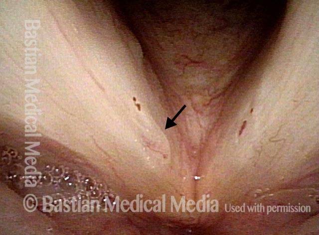
Three weeks later, the vocal cords now match, voice is improved, and the site of surgery (arrow) is healed.

Sfumature “raccolte” dagli esami quotidiani

Visual Portfolio, Posts & Image Gallery for WordPress

Vocal “overdoer” (1 of 4)

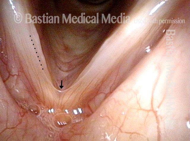
A vocal “overdoer” with hoarseness. Note broad-based swelling of both vocal cord margins. A microweb, thought to be congenital, is also seen at the arrow. Tiny dots indicate a subtle wrinkle or shallow sulcus.

Inspiratory phonation (2 of 4)

The patient has been asked to produce inspiratory phonation to reveal the translucent polyp and “sulcus,” again at tiny dots.

Translucent polyp (3 of 4)

Under strobe light, the translucence of the polypoid elevation is seen more clearly.

Open phase (4 of 4)

At open phase of vibration again under strobe light, the broad based left vocal cord elevation is also seen.

L’espressione della lesione nella mucosa varia

Visual Portfolio, Posts & Image Gallery for WordPress

Vocal cord injuries (1 of 4)

Vocal cord injuries of overuse are often bilaterally similar, but here we have two quite different expressions of injury: fibrosis and capillary ectasia on left (right of photo); translucent polypoid injury (not a cyst) on the right (left of photo).

Narrow band lighting (2 of 4)

Now under narrow band light, the left cord (right of picture) has a flatter, fibrotic expression with tiny ectatic capillaries.

Strobe lighting (3 of 4)

Under strobe light, the translucent, polypoid nodule of the right cord (left of photo) distorts vibratory closure.

Phonation (4 of 4)

This is the best closure this grossly hoarse person can achieve.

Il potere di vedere un polipo “vicino-chiaro” e non “lontano-sfocato”.

Visual Portfolio, Posts & Image Gallery for WordPress

Disant view (1 of 4)

This man is a singer. Distant “far” examination shows what appears to be a subtle or even inconsequential elevation of his right vocal cord (left of photo).

Closer view (2 of 4)

Closer view shows the lesion better, but its true magnitude is about to be seen…when a truly close-clear view is achieved.

Close-clear view (3 of 4)

Close-clear view under strobe light , falsetto voice, closed phase of vibration. The broad-based swelling is more clearly seen, between the two dots.

Open phase (4 of 4)

Open phase of vibration again shows the injury.

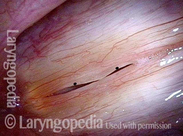
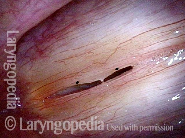
Polipo o cisti?

Visual Portfolio, Posts & Image Gallery for WordPress

Hoarseness (1 of 4)

During an upper respiratory infection, this older woman developed hoarseness that has not gone away during the past year. Is this the end stage perhaps of a hemorrhagic polyp?

Position of lesion (2 of 4)

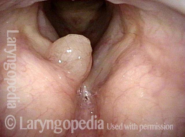
In this slightly closer view, with the patient breathing out, the lesion appears too “high” within the laryngeal vestibule, and not truly at the level of the vocal cords.

Close view (3 of 4)

This close view is on the way to determining if there is any attachment to the vocal cords themselves. Not quite yet able to tell…

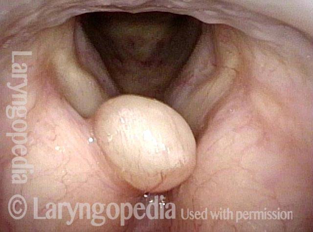
Anterior saccular cyst (4 of 4)

The tip of the scope has just passed the lesion and the vocal cords are unaffected. As it appears to be arising from the ventricle, it could be classified as an anterior saccular cyst. Likely the saccule or a mucus gland became plugged due to inflammation during the upper respiratory infection a year earlier, and it filled with mucus.

Il piccolo segmento vibrante dà la voce di un piccolo fischio di latta

Visual Portfolio, Posts & Image Gallery for WordPress

Prephonatory instant (1 of 6)

This young woman has been hoarse for many years. This preparatory posture shows marked separation of the cords posteriorly, suggesting MTD as well.

Phonation (2 of 6)

Now producing voice, with vibratory blur of the entire length of the cords on both sides.

Gaps due to nodules (3 of 6)

Under strobe light at a lower pitch of A4 (440 Hz), closed phase of vibration. Large gaps anterior and posterior to the polypoid nodule(s) explain breathy quality and short phonation time.

Open phase (4 of 6)

Open phase of vibration also at A4 (440 Hz) shows that the full length of the vocal cords are vibrating. Compare with the following two photos.

“Tin whistle” sound (5 of 6)

Now at A5 (880 Hz), the patient can only make an extremely tiny (tin whistle) quality. The only segment vibrating is within the circle (here, closed phase). The posterior segment does not vibrate.

“Tin whistle” at open vibration (6 of 6)

Still at A5 (880 Hz), the open phase of vibration, again of *only* the tiny anterior segment.

La riduzione dei polipi del fumatore migliora la voce anche se il risultato della laringe potrebbe non essere “carino”

Visual Portfolio, Posts & Image Gallery for WordPress

Smokers Polyp (1 of 5)

Six years after vocal polyp removal elsewhere. As a result of continued smoking, the voice is deep and rough, and an obvious recurrent/residual “smoker’s” polyp is seen on the right vocal cord(arrow, left of photo). The patient “hates” her rough and masculine voice quality.

Reine’s edema (2 of 5)

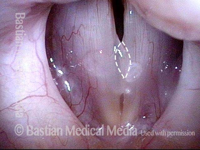
Inspiratory phonation is elicited to “pull” the redundant tissue medially, revealing a lot of Reine’s edema of the left vocal cord, too. The dotted lines show the ellipse of mucosa that will be removed during surgery. Mucose will be preserved at the margins and the gelatinous lateral within the polyps will be suctioned away if liquid, and dissected away if fibrotic.

A week after surgery (3 of 5)

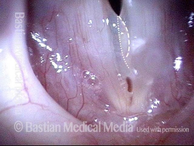
The dotted lines show the extent of mucosal excision—an area that will take a few weeks to re-mucosalize. Since this was a polyp “reduction,” though hoarse this early post, she has a ” functional” voice. There should be no alarm if patients are aphonic for a week or even a few weeks while inflammation resolves.

Residual Reinke’s edema (4 of 5)

At two months post, the patient is very pleased and says the improvement to voice is “large.” Here, it appears there may be some residual Reinke’s edema especially of the left vocal cord (right of photo) judging by the slightly convex margin.

Residual submucosal edema (5 of 5)

With inspiratory phonation, the residual submucosal edema is made obvious, especially on the left. This was (as intended) a polyp “reduction” approach, rather than polyp “removal” as the latter is too hard on voice, and it is not possible to “put back” if too much tissue is taken. Here, there is no stiffness, and if desired, more can be removed. Of course, since the patient is so pleased with her voice, no further treatment is needed.
Vocal nodules & other voice injuries YT Thumbnail
Riproduci video

Noduli e altre lesioni delle corde vocali: come si verificano e come possono essere trattati

Questo video spiega come si verificano i noduli e altre lesioni delle corde vocali: attraverso l’eccessiva vibrazione delle corde vocali, che si verifica con un uso eccessivo della voce.

Dopo aver posto queste basi, il video prosegue esplorando il ruolo delle opzioni terapeutiche come la terapia vocale e la microchirurgia delle corde vocali.

Esempio audio 1

Commenti dei pazienti sul miglioramento della voce dopo la rimozione chirurgica di un polipo delle corde vocali:

Esempio audio 2

Qualità della voce, con un polipo vocale, PRIMA dell’intervento chirurgico:

Stesso paziente, DOPO l’intervento chirurgico:
Videoconsulto con
il dottor Bastian
Hai ancora domande? Pianifica una chiamata con il Dr. Bastian.

Table of Contents

Subscribe
Notify of

0 Comments
Newest
Oldest Most Voted
Inline Feedbacks
View all comments
0
Click to see all comments.x