An encyclopedia about voice, swallowing, airway, coughing, & other head + neck disorders.

Phonatory insufficiency refers to when the vocal cords cannot close sufficiently or vibrate adequately to produce a serviceable voice. An inability to close is usually evidenced by air-wasting phenomenology.

This phonatory insufficiency could have one of several causes. It could be due to the loss of part or all of one or both vocal cords, such as after removal of a vocal cord cancer. Or it could follow prolonged intubation and resulting pressure necrosis of the posterior ends of the vocal cords 1. Another possibility might be scarring of the anterior joint capsule of the cricoarytenoid joints, also as a complication of prolonged endotracheal intubation due to grave illness. Yet another cause might be vocal cord paralysis or paresis. The latter problems not only interfere with the cords’ ability to close, but also make the affected cord flaccid, so that it blows out of the way too easily, further wasting the air stream.

When a person with any of these causes of poor vocal cord closure tries to produce voice, maximum phonation time is typically reduced, because only a fraction of the air pushed up from the lungs is converted to sound, with the remainder of the air quickly “wasted.”

The second main category of phonatory insufficiency, in which the vocal cords cannot vibrate adequately, is seen in a person with stiff or scarred vocal cords. Such a person may not waste air, but just be unable to produce other than a harsh whispery sound, because the stiffened vocal cords (now more like thick leather rather than like, as is normal, plastic wrap overlying a thin layer of jello) cannot vibrate as freely or at all.


Photos:

Visual Portfolio, Posts & Image Gallery for WordPress

Phonatory insufficiency (1 of 3)

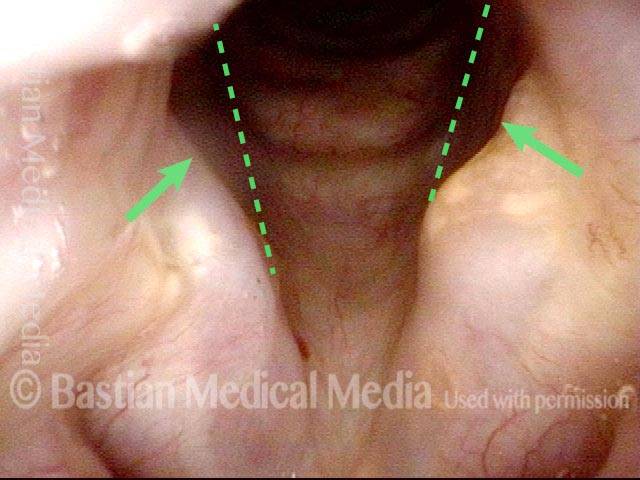
Abducted breathing position. Note the divots at the posterior commissure (arrows), likely due to pressure necrosis caused by intubation of long duration. Dotted lines indicate the lines of the normal cord, to show the divots more clearly.

Phonatory insufficiency (2 of 3)

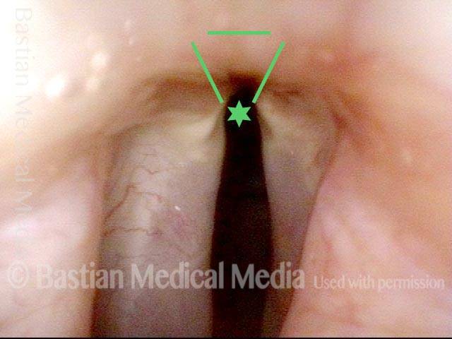
The irregular white line along the length of the vocal cords (arrows) suggests that there may have been pressure necrosis of the musculo-membranous portion of the vocal cord and that now the mucosa adheres directly to muscle, with no intervening vocal ligament layer.

Phonatory insufficiency (3 of 3)

Maximum phonatory closure. Note that the posterior commissure defect is hidden by the partial closure of the arytenoid cartilages. Even so, the arytenoid cartilages are unable to come into contact. The musculomembranous cords are quite far apart due partly to tissue loss. Furthermore, the cords are stiff and inflexible. No glottic voice is possible.

Example 2

Visual Portfolio, Posts & Image Gallery for WordPress

Phonatory insufficiency (1 of 4)

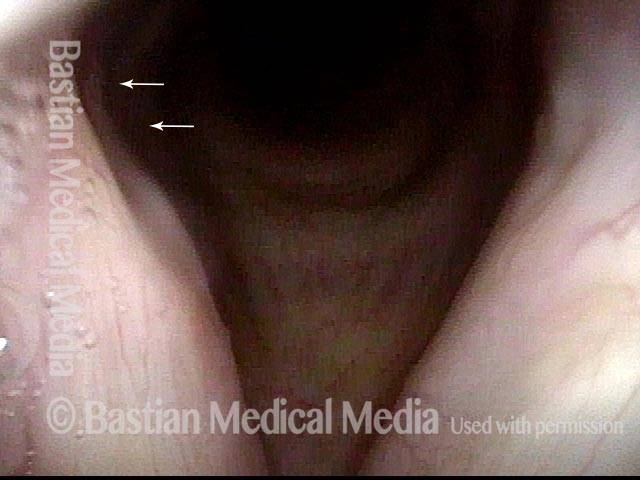
After 15 days of intubation, this voice is sounding both breathy (air-wasting) and pressed. From a distance it appears that the right cord (left of image) is paralyzed. (Compare with image 2)

Phonatory insufficiency (2 of 4)

During phonation, the voice again sounds breathy and pressed.

Phonatory insufficiency (3 of 4)

A close up view shows the posterior divot of the right cord (left of image). The absence of atrophy, bowing, or flaccidity, confirms that the problem is right cord fixation due to scarring of the right cricoarytenoid joint, not paralysis.

Phonatory insufficiency (4 of 4)

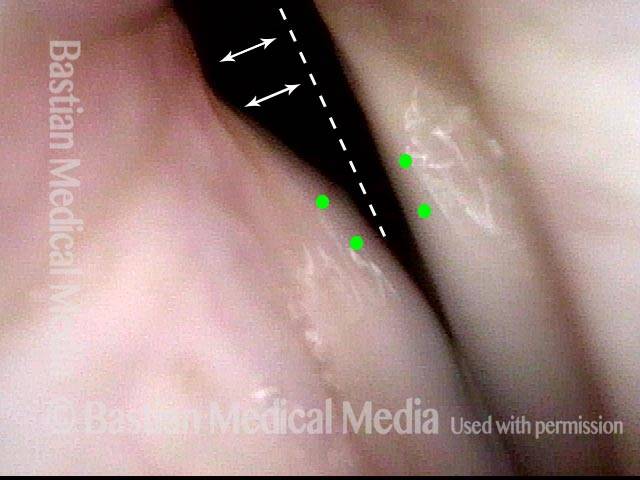
During phonation, the posterior commissure deficit caused by pressure necrosis from the endotracheal tube is seen with the dotted line. The small green circles represent the vocal processes not approximating thus validating the joint injury.

Phonatory Insufficiency Due to Loss of Vibration-capable Tissue

Visual Portfolio, Posts & Image Gallery for WordPress

Phonatory insufficiency due to loss of vibration-capable tissue (1 of 4)

The larynx is in an open position for breathing, though the right half of the larynx has been removed to treat cancer. On the right (left of photo) there is a pseudocord, which is tissue used to replace the right cord, but not a type of tissue that can vibrate. Only the middle of the left cord (right of photo) has been preserved.

Phonatory insufficiency due to loss of vibration-capable tissue (2 of 4)

Here the patient produces voice by bringing the remnant of the left cord (right of photo) into partial contact with the pseudocord (left of photo) . You can see vibratory blurring of the left cord, but no blurring of the pseudocord. In addition, closure is incomplete.

Phonatory insufficiency due to loss of vibration-capable tissue (3 of 4)

This image shows voice use under strobe lighting. During the closed phase of vibration the cords do not fully close as the left cord cannot quite reach the pseudocord. This accounts for air-wasting.

Phonatory insufficiency due to loss of vibration-capable tissue (4 of 4)

Still under strobe lighting, this image shows the open stage of vibration. The pseudocord appears the same, but the remnant of left cord has oscillated laterally. Due to the shortened length of the cord, the pitch of the voice is higher than expected for this person.
  1. Bastian RW, Richardson BE. Postintubation phonatory insufficiency: an elusive diagnosis. Otolaryngol Head and Neck Surg. 2001; 124(6): 625-33. []