Tracheoesophageal Party Wall with Wheezing
Abducted breathing position (1 of 5)
Normal laryngeal entrance, with vocal cords in abducted (breathing) position.
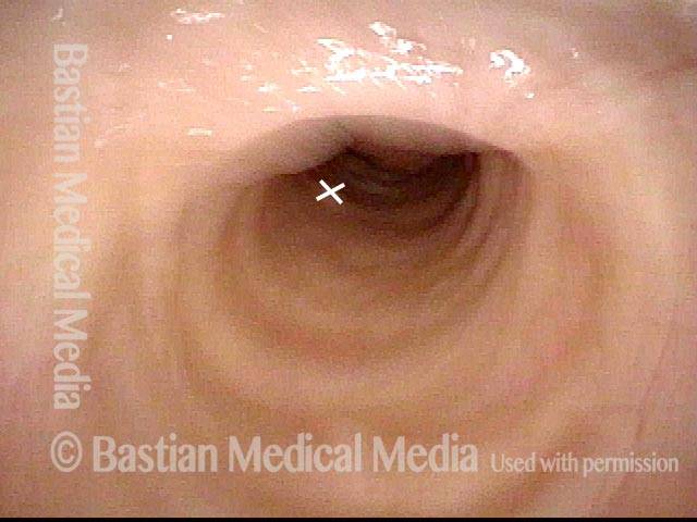
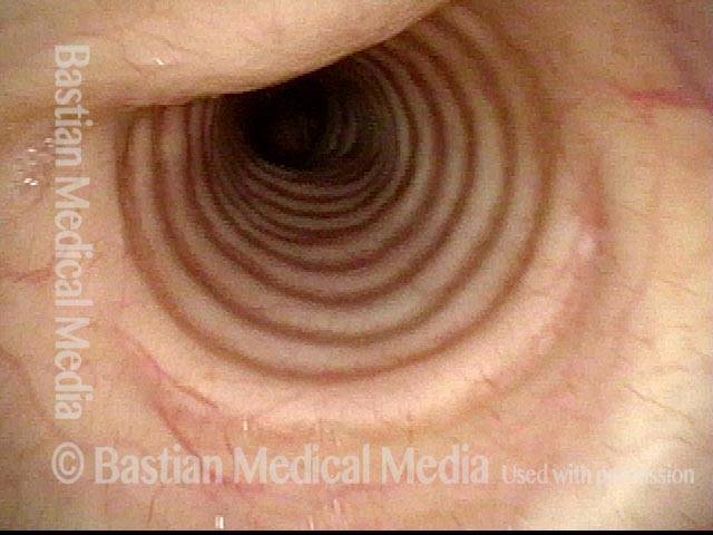
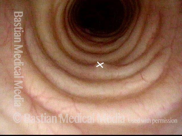
View of mid-trachea (2 of 5)
View in normal mid-trachea. Note that the cartilaginous rings make up approximately 2/3 of the circumference and that the membranous trachea (upper photo at ‘X’) is more flat.
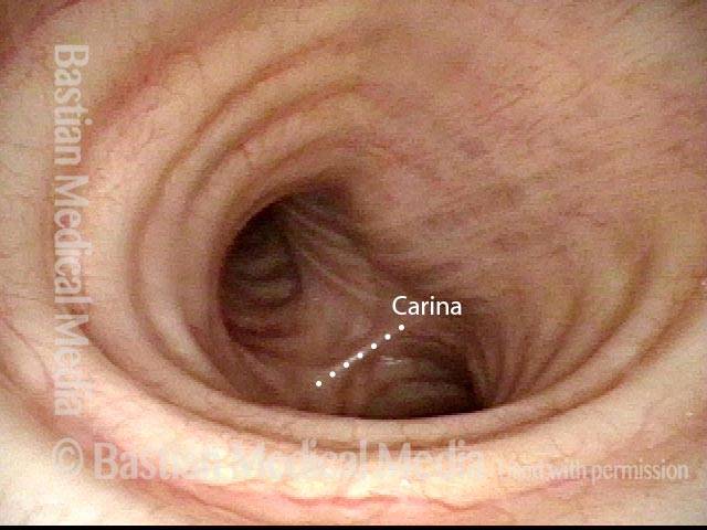
View just above the carina (3 of 5)
View just above the carina, where the distal trachea splits into left and right mainstem bronchi. Anterior take-off of carina at the ‘X’. The straight line delimits the membranous (flexible) tracheal wall.
Wheezing begins (4 of 5)
With Valsalva maneuver to accentuate patient’s functional expiratory wheezing. Note that the membranous tracheal and bronchial walls bulge inward on a functional basis to narrow the airway. Wheezing begins to be heard. The ‘X’ again marks the anterior take-off of the carina. Compare with Photo 3.
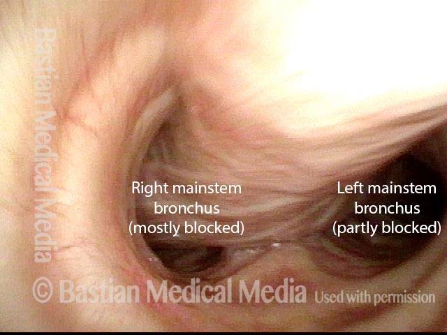
Left bronchus blocked (5 of 5)
As bulging inward continues, the left mainstem bronchus is particularly blocked. This explains why, on auscultation of the chest, wheezing sounds louder on the left than the right. Compare with photos 3 and 4.
Upper Airway Wheezing, as a Kind of “Skill”
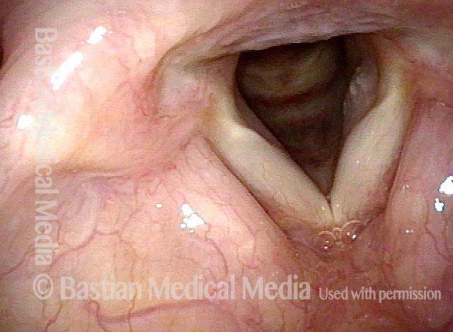
At the level of the vocal cords (1 of 8)
Vocal cords, in breathing position, in a person with wheezing who has been diagnosed elsewhere with asthma. Nothing seen here to explain the wheezing.
In the upper trachea: Valsalva maneuver (2 of 8)
Now descended into the upper trachea. As the patient performs (upon request) a semi-Valsalva manuever, one can see mild inward bulging of the membranous trachea (top- left of photo). This is not sufficient to explain the patient’s wheezing.
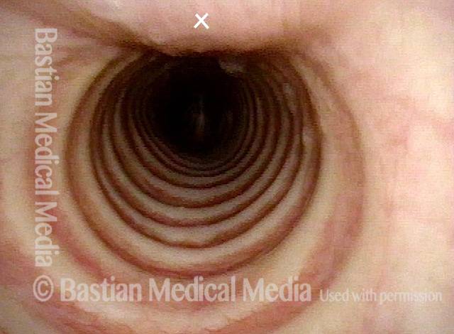
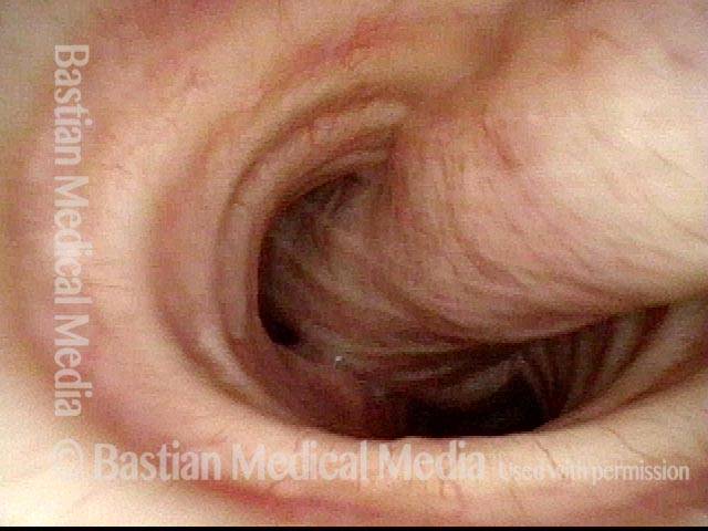
In the mid-trachea: quiet breathing (3 of 8)
Descended further, into the mid-trachea, with the carina (dotted line) seen in the distance, as the patient breathes quietly.
In the mid-trachea: Valsalva maneuver (3 of 8)
Same view as photo 3, but during another semi-Valsalva maneuver. The membranous tracheal wall bulges inward again, but much more noticeably here than in the upper trachea (photo 2), especially down by the carina (which is now hidden from view).
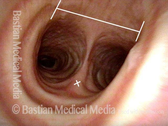
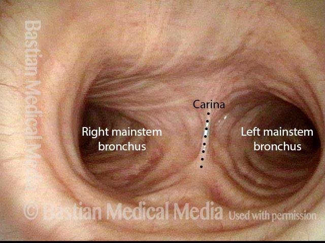
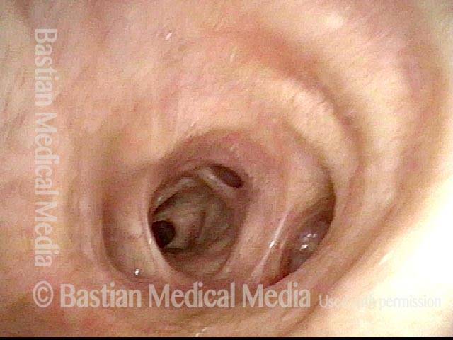
In the lower trachea: quiet breathing (5 of 8)
Down further yet, almost to the level of the carina (dotted line). On each side is the entrance to each of the mainstream bronchi.
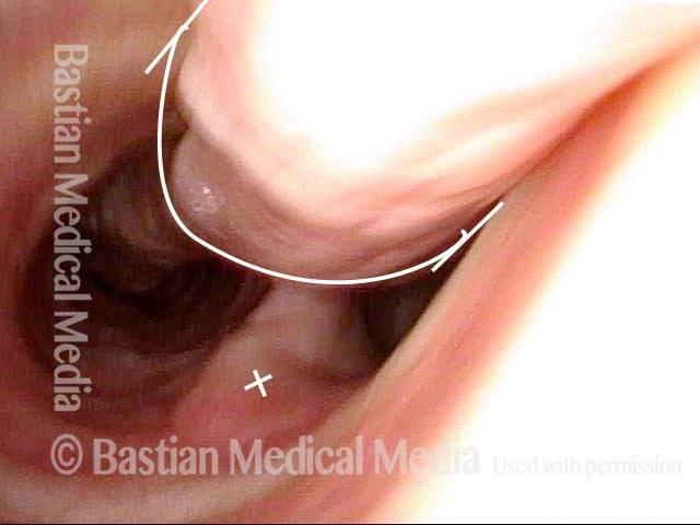
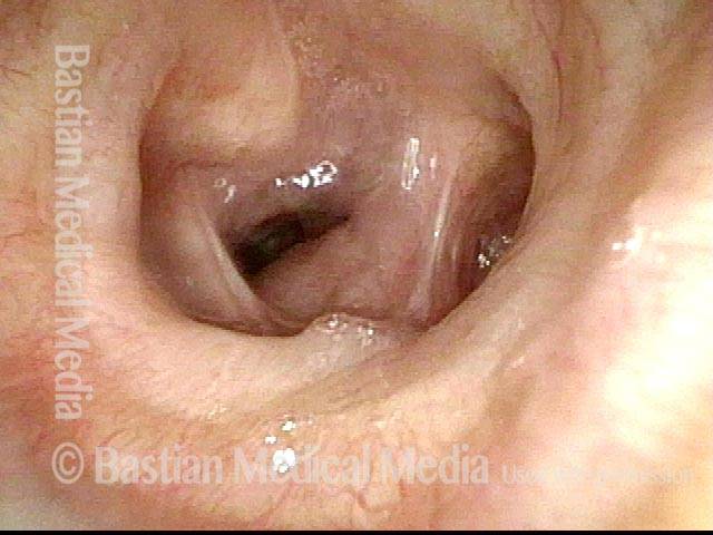
In the lower trachea: Valsalva maneuver (6 of 8)
Same view as photo 5, but during another semi-Valsalva maneuver. The membranous tracheal wall bulges inward again to partly block the airway, especially the right mainstem bronchus. This patient’s wheezing sounds loudest over the sternal notch and manubrium; softer wheezing is also heard in the distal lung fields, and as expected from this photo, is louder on the right than the left.
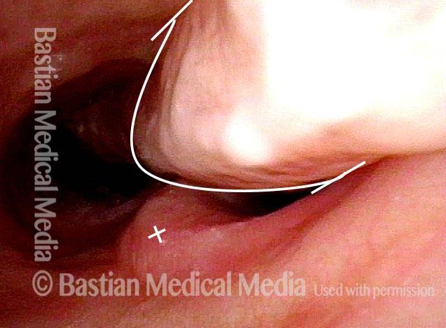
In the right mainstem bronchus: quiet breathing (7 of 8)
Further down yet, now looking directly into the right mainstem bronchus.
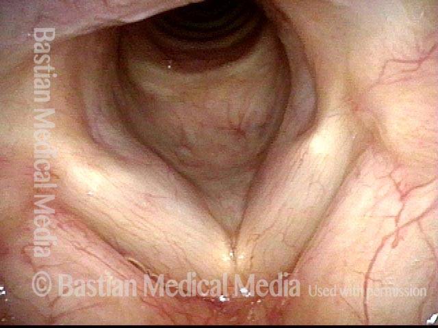
In the right mainstem bronchus: Valsalva maneuver (8 of 8)
Same view as photo 7, but during another semi-Valsalva maneuver. Note compression as well of lobar bronchi, also partly responsible for the patient’s wheezing.
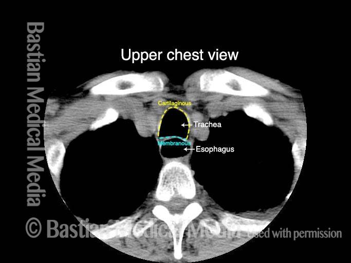
Rumbling Vibration of the Tracheoesophageal Party Wall can make Coughing Sound “Infectious and Productive” when it isn’t
Tracheobronchial cough vibration (1 of 2)
Patient with SNC who is treated for presumed infection because of her congested rumbling “productive-sounding” cough. The “X” marks the same place in this photo and the following photo.
Tracheobronchial cough vibration (2 of 2)
Patient is at the moment of a deep, productive-sounding cough, but in fact it is not productive. Her many courses of antibiotics are probably unnecessary. The narrowing of the lumen is due to inward bulging of the membranous tracheal wall. The blur is caused by vibration during this cough.