Common Tests in Laryngology: A Patient Guide (Alphabetized)
Patients often ask what various tests involve in Laryngology. Here, in layperson’s terms, are brief explanations of common studies you might be asked to undergo for voice and swallowing evaluation:
Barium Swallow
You drink a radiopaque (X-ray visible) barium solution, which outlines the esophagus and stomach. It assesses:
- Transit speed
- Hiatal hernia
- Tumors
- Abnormal contractions
- Acid reflux
Note: This test is not ideal for evaluating swallowing disorders.

Barium Swallow (Barium Esophagram: Anterior-Posterior View)
This video presents a clear visual example of a barium swallow, a test that involves having the patient swallow a barium solution while using x-rays to observe the flow of the barium, which can reveal swallowing deficiencies.
CXR (Chest X-ray)
A basic X-ray of the chest, usually done in two views:
It’s a quick, inexpensive way to screen for lung, heart, or rib abnormalities.

CT Scan or CAT Scan (Computed Tomography)
These terms refer to the same test. A CT scan uses many low-dose X-ray images, compiled by computer into “slices” of the body.
These reconstructions can be:
- Axial (horizontal) – like slicing a loaf of bread from top to bottom
- Coronal (front to back) – from nose to back of head
- Sagittal (side to side) – from ear to ear
CT scans can be done with or without contrast, an intravenous dye that highlights blood vessels and lymph nodes. This test is often preferred for evaluating structures like the larynx.

EKG or ECG (Electrocardiogram)
This test evaluates the electrical activity of the heart. Sticky pads are placed on the chest, and wires connect to a monitor that records heart rhythms. It can detect signs of:
- Heart attacks (past or current)
- Arrhythmias (irregular heartbeats)
- Heart failure
(Note: “EKG” uses the German spelling with a “K.”)
EMG (Electromyography)
This test measures the electrical signals of muscles. Sticky pads (ground and reference) are used, and then a hollow, Teflon-coated needle electrode (insulated except at the tip) is inserted into a muscle to assess how it functions at rest and during use.
In laryngology, EMG may assess muscles controlling the vocal cords, or to verify accurate placement before injecting botulinum toxin (“botox”) into vocal cord muscles for treatment of spasmodic dysphonia, or the upper esophageal sphincter (cricopharyngeus muscle) to treat retrograde cricopharyngeus dysfunction (R-CPD).

MRI (Magnetic Resonance Imaging)
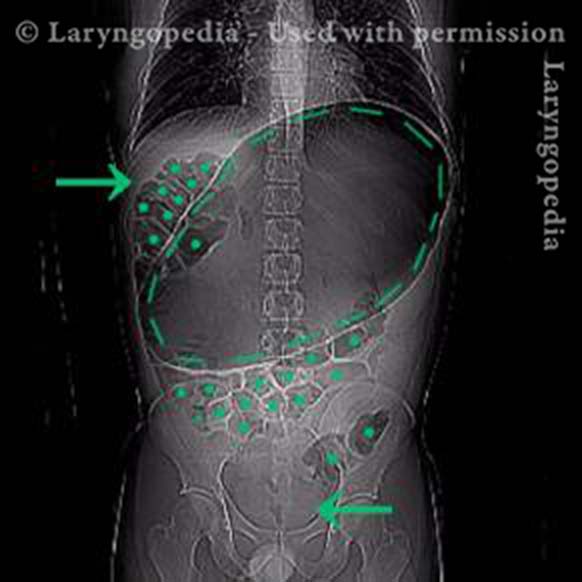
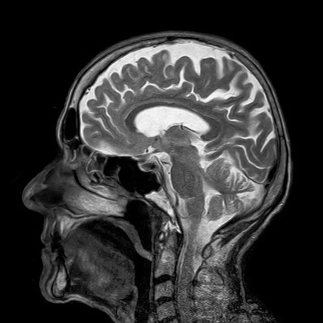
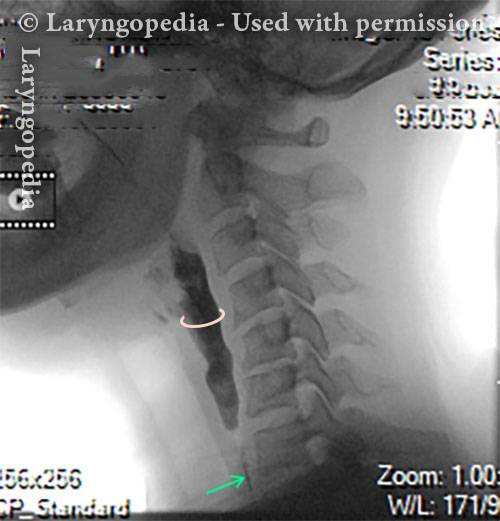
An MRI does not use X-rays. Instead, it employs magnetic energy to change the polarity of atoms in the body, which are then translated into detailed images by computer. Like CT, MRI can produce axial, coronal, and sagittal images. MRI is often superior for viewing soft tissues, spinal cord, brain, and nerves.

PET Scan (Positron Emission Tomography)
PET is a functional imaging test, highlighting areas of high metabolic activity—often associated with cancer. A radioactively tagged sugar is injected, and cancerous areas absorb more of it, appearing as “hot spots.” Because PET lacks clear anatomical detail, images are typically fused with a CT scan (called PET-CT) for precise localization.
Videofluoroscopic Swallow Study (VFSS)
Also called a Modified Barium Swallow or “Cookie Swallow,” this is the gold standard for evaluating swallowing.
Barium is given in small amounts by spoon (liquid and puree) and as paste on a cookie. X-ray images show:
- How material moves from the mouth to the esophagus
- Whether residue remains in the throat
- If barium enters the larynx (penetration) or lungs (aspiration)
- Whether the upper esophageal sphincter relaxes properly

Videostroboscopy (aka Strobovideolaryngoscopy)
This test evaluates vocal cord anatomy and vibration. It includes:
- Voice testing and recording
- Flexible or rigid scope examination of the larynx, subglottis, and trachea
- Strobe light imaging of vocal cord vibration
The session is recorded for review with the patient, referring doctor, speech pathologist, or other professionals.

Share this article