An encyclopedia about voice, swallowing, airway, coughing, & other head + neck disorders.

The Red Herring Pitfall in Diagnosing Voice, Swallowing, Airway, Cough, and Burping Disorders

Robert W. Bastian, M.D. — Published: February 18, 2025

The Origin of the Term “Red Herring”

The phrase “red herring” originates from the use of hunting dogs in Scandinavia and England. In training mode, a fox pelt might be dragged through the forest to lay down the scent. When the dogs became proficient, the trainers would increase the difficulty. Smoked herrings, which turn a deep reddish color and emit a strong odor, might be dragged across the “scent trail” of the fox to distract and mislead the dogs.

Over time, this exercise taught dogs to stay focused on their intended quarry rather than being distracted by a false lead. The term evolved into a metaphor for any misleading clue or distraction that diverts attention from the truth, the main idea, or the subject.

Red Herrings in the Diagnostic Process and Protection Provided by the Integrative Diagnostic Model

By analogy, red herrings can distract clinicians trying to “follow the trail” leading to an accurate diagnosis.

To protect against this kind of potential error, the integrative diagnostic model (described elsewhere) excels.

This model ensures that patient history, the vocal capability battery, and an intense examination serve as a “three-legged stool” that stands firmly when each component is utilized fully. In integrating these three sources of information, each serves as a double-check against the other two.

Examples:

1) Red Herrings in the Patient History

A patient who suffers from respiratory allergies might attribute voice changes to that condition. The clinician may be protected from this “red herring” by learning additional information: the patient rates a “7 and 7” on the talkativeness and vocal loudness scales, is a kindergarten teacher, has three children under six at home, and an elderly parent with hearing loss.

Here, a careful history prevents the “red herring” of allergy from obscuring the true issue: vibratory injury of the vocal cords from overuse. Subsequent “swelling checks” in the vocal capability battery and high-quality examination further clarify the diagnosis by revealing the vocal phenomenology and visual findings of vocal nodules.

2) Red Herrings in the Vocal Capability Battery

A patient’s initially normal-sounding speaking voice may be a “red herring” that predisposes the clinician to assume normal function. Yet, during vocal capability elicitation, subtle but definite jittery instability on sustained phonation and rare “catches” at low pitch phonation may protect the clinician from this assumption when the actual diagnosis is mild spasmodic dysphonia.

Similarly, a patient who initially sounds like full-blown spasmodic dysphonia may report repeated episodes. Recognizing that spasmodic dysphonia is virtually always an unremitting chronic voice change (though variable in severity moment-to-moment) protects against a red herring. Further rapid-fire elicitation of varied vocal tasks may confirm that the voice disorder is actually non-organic or malingering.

3) Red Herrings in the Examination

The physical and endoscopic examination is another area where misleading findings can arise. Leukoplakia in a young singer may mislead the clinician toward a diagnosis of reflux. However, the patient’s absolute lack of reflux symptomatology (despite the possibility of “silent” reflux), non-response to aggressive reflux management, and, most importantly, findings of faint vascular stippling seen only under narrow-band light at close range, lead instead to a correct diagnosis: HPV 16 infection.

Often, “normal” is the red herring examination result. Without an intense examination, telltale findings of intubation injury may go unnoticed, or subtle paresis may be overlooked.

The Importance of Clinical Reasoning and Synthesis

Avoiding diagnostic red herrings requires synthesis. Clinicians must integrate patient history, vocal capability evaluation of vocal phenomenology, and intense, close-range, highly magnified examination. Remaining attuned to each of the three sources of information and ensuring coherence within and among them protects against premature conclusions based on red herrings.

Ideally, one clinician (physician or speech pathologist) will have mastered all three parts of the integrative model, since the integrative process is far more efficient within one brain, than between two. Less ideally, these components may be distributed between a physician and a speech pathologist.

Where the integrative diagnostic model is mastered, so-called "objective measures" serve only for documentation and possibly biofeedback, but not for diagnosis.

Leukoplakia as a Visual Red Herring

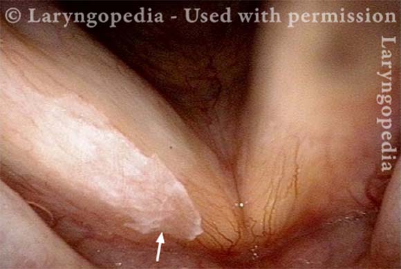
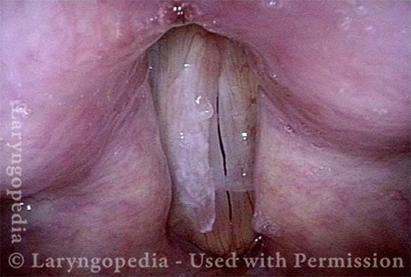
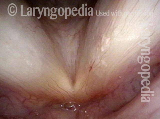
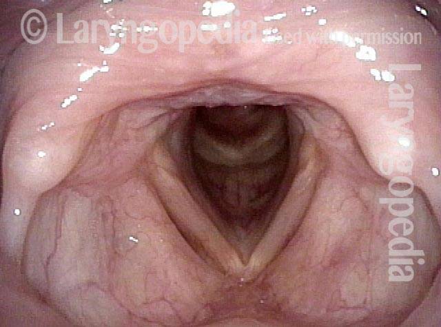
This middle-aged man came for evaluation and treatment of a “white lesion” that his doctors was seen elsewhere on a vocal cord, which they thought explained voice change. He had experienced that voice change suddenly a few months before (Red herring alert, as he was not a smoker, and white lesions do not usually appear and cause voice change abruptly).

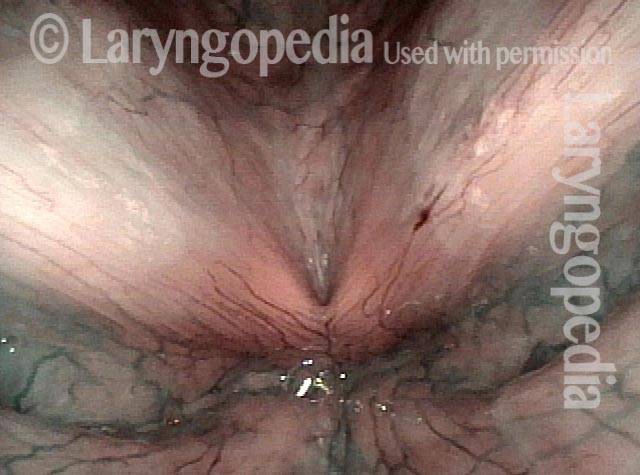
Furthermore, his main vocal issue was not so much hoarseness, as a tendency for the voice to fade with use across the day. This is not the usual phenomenology of leukoplakia, which would be expected to produce the same voice from morning to night.  As it turns out, the leukoplakia was incidental to his voice change, which was instead due to scissoring / overlap of one cord on the other and bowing/flaccidity—possible TA-only paresis of the right cord.

Visual Portfolio, Posts & Image Gallery for WordPress

The Red Herring Lesion (1 of 4)

At close range, the white patch on the upper surface of the right cord (left of photo) is closer to “spilled milk” than “cake icing.” And it doesn’t extend to the vocal cord margin. This means it is unlikely to interfere much with vibration or match of the two cords during phonation.

Bowing and scissoring (2 of 4)

At the prephonatory instant, the overlap of right cord (left of photo) over top of the left (dotted lines). Also the leukoplakia does not interfere with match between bowed vocal cord margins, right greater than left.

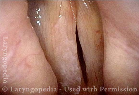
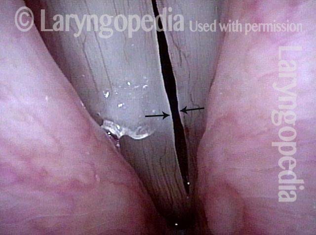
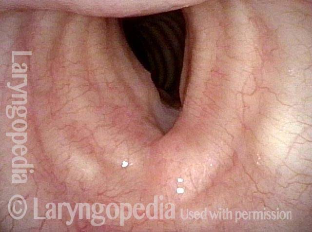
Strobe light, open phase of vibration (3 of 4)

Here, the bowing of right cord as compared with the left is seen more clearly, suggesting TA-only paresis. That diagnosis would be more coherent with abrupt voice change, than leukoplakia would.

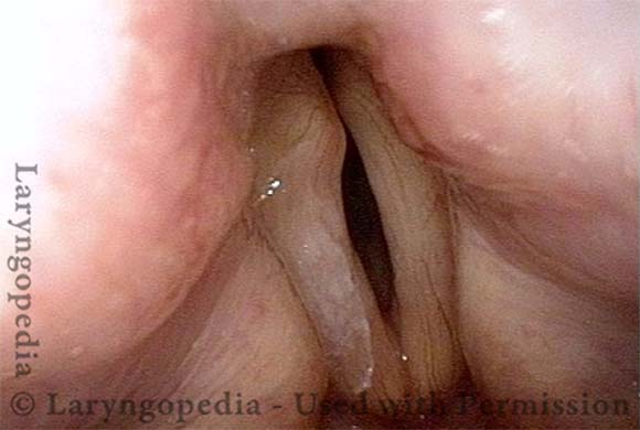
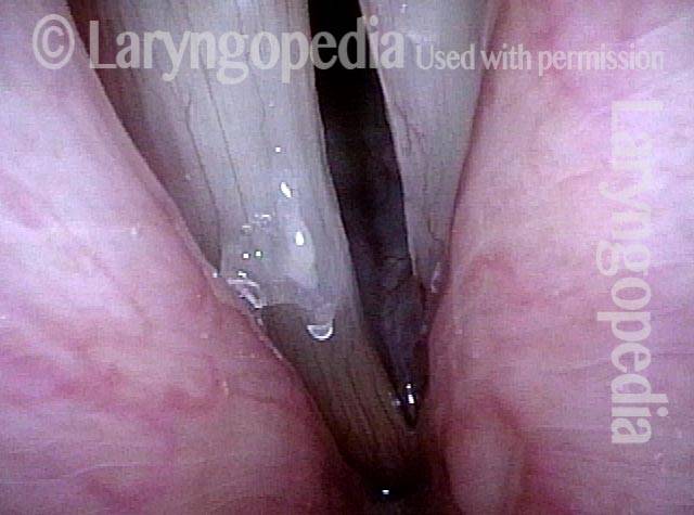
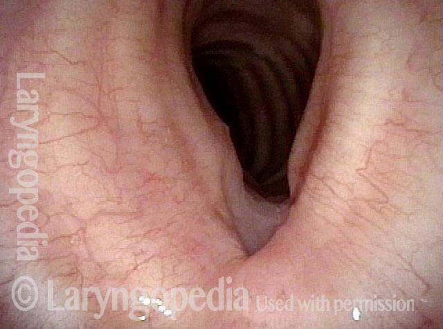
Closed phase of vibration (4 of 4)

There is mismatch of levels with right cord higher than the left, but no margin effect from the leukoplakia. As an explanation for this man’s voice change, his leukoplakia is a red herring!

Pushing Past Red Herrings to Find the Real Issue

Visual Portfolio, Posts & Image Gallery for WordPress

Obvious lesion not important (1 of 3)
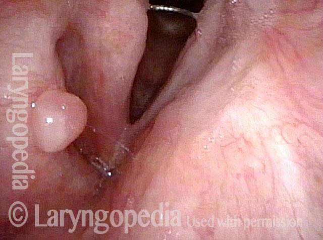
Several months after removal of exuberant papillomas, voice remains quite good, but is becoming a little deeper. The obvious lesion here is not important; the subtle one is the key.

Granuloma (2 of 3)

Narrow band light reveals the spherical lesion to be a granuloma, not papilloma (which would have stippled vascular markings).

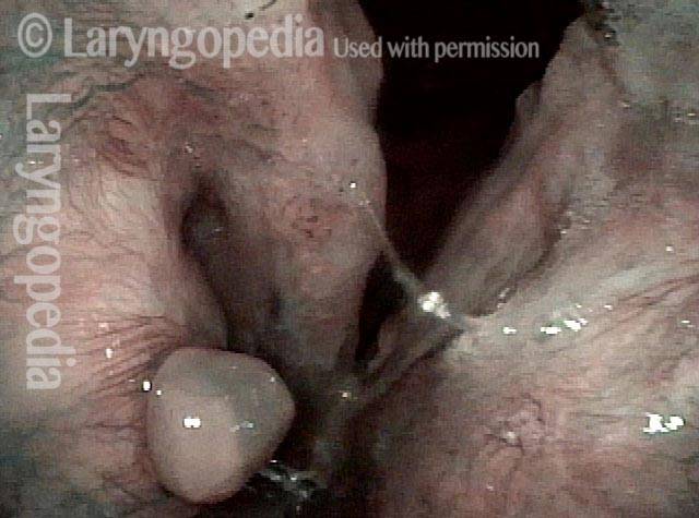
Carpet-varient papilloma (3 of 3)

At closer range, still under narrow band light, carpet-variant papilloma can be seen on the posterior right vocal cord (left of photo). This is the important finding.

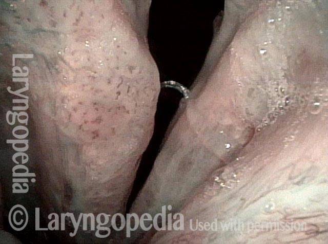
Red Herring Capillary Ectasia and Mucosal Injuries

Visual Portfolio, Posts & Image Gallery for WordPress

Ectatic capillary (1 of 4)

This young performer has a sense of a weakened voice and loss of vocal stamina. Here, we see an ectatic capillary of the left vocal cord (right of photo). Is the problem intermittent vocal hemorrhage from this vulnerable capillary? Is there increased susceptibility to edema due to this margin capillary?

Ectatic capillary, narrow band light (2 of 4)

Under narrow band light, the capillary is even more evident. The additional network of prominent capillaries prompt the same questions as in caption 1.

Margin swelling (3 of 4)

Under strobe light at B-flat 4 (494 Hz), we see subtle margin swelling (arrows), here of only “indicator lesion” magnitude.

Bowing, atrophy, and flaccidity (4 of 4)

The large amplitude of the open phase of vibration at the same pitch, along with the lack of closure in photo 3, reveals the actual problem to be bowing, atrophy, and flaccidity. These findings fit with the “bowing” symptom complex: loss of edge to voice quality and the tendency of voice quality and strength to “fade” as the day progresses.

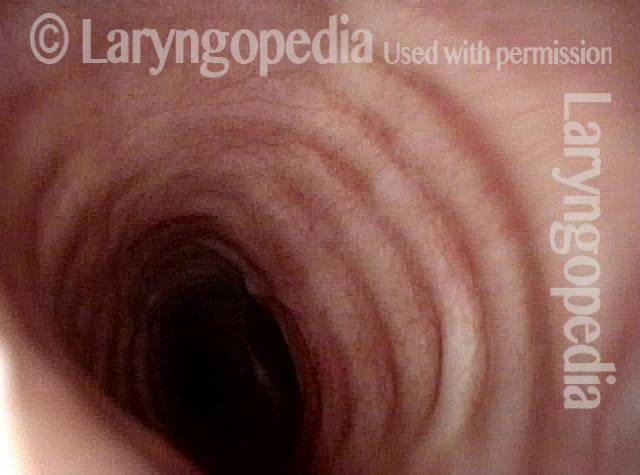
Tracheal Stenosis Here Is A Red Herring; the Diagnosis Is Actually CPS

Visual Portfolio, Posts & Image Gallery for WordPress

Tracheal stenosis? (1 of 4)

During a grave illness, this woman eventually underwent tracheotomy. Though she wore the tube for several months, it was removed 3 years ago. Only six months prior to this examination, due to a feeling of choking, she underwent a CT scan that revealed tracheal stenosis. The patient does have mildly noisy breathing but has no sense of exercise intolerance.

Narrowing at trachea (2 of 4)

Viewing from just below the vocal cords, there is narrowing and deformity of the trachea at the site of prior tracheotomy.

Closer view (3 of 4)

A closer view shows normal trachea beyond.

No significant change in breathing (4 of 4)

Now with the scope through the area of greatest narrowing, the patient doesn’t experience any significant change in her breathing. Her symptoms are those of cricopharyngeus spasm, not tracheal stenosis.
Subscribe
Notify of

0 Comments
Newest
Oldest Most Voted
Inline Feedbacks
View all comments
0
Click to see all comments.x