Laryngocele
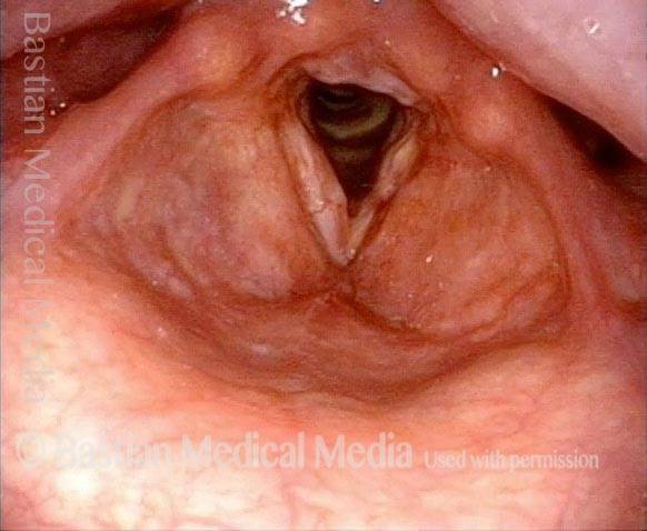
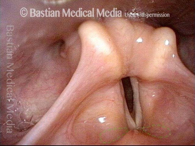
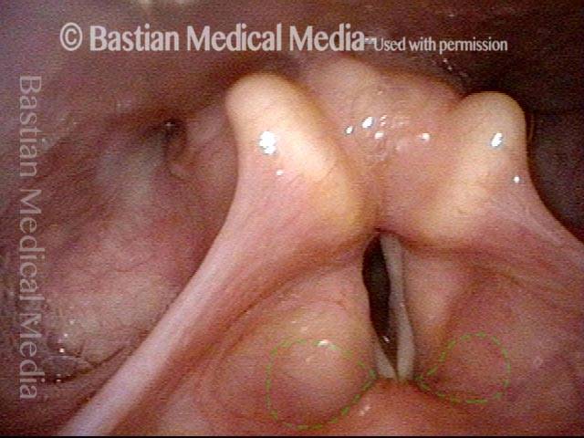
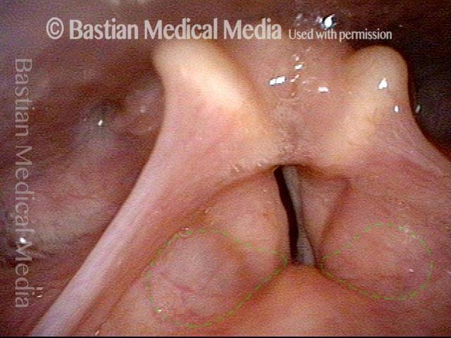
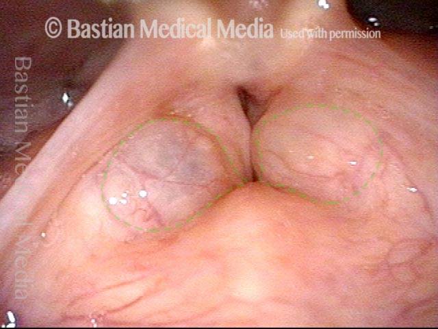
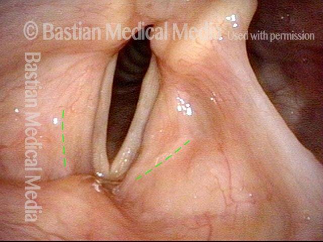
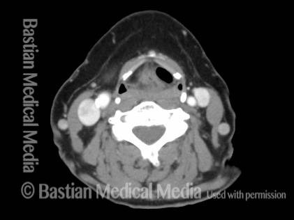
Laryngocele is a disorder in which the laryngeal saccule is inflated and becomes abnormally enlarged. A common symptom of a laryngocele is hoarseness.
How It Develops
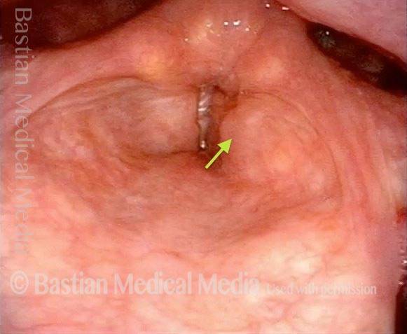
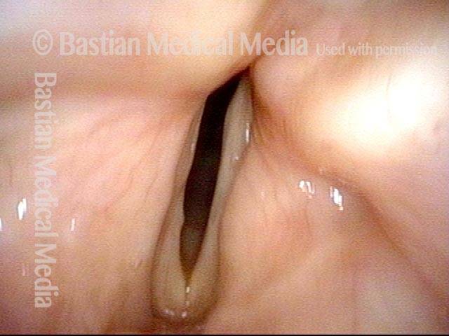
The laryngeal saccule, or laryngeal appendix, is a very small blind sac—a dead-end corridor, so to speak—which is located just above the vocal cords, one on each side, and is lined with glands that supply lubrication to the cords. When a person makes voice, it is possible for a little bit of the air being pushed up out of the trachea to slip into this saccule. If over time enough air enters the saccule with enough force, the saccule may begin to be inflated and stretched out, leading to a laryngocele.
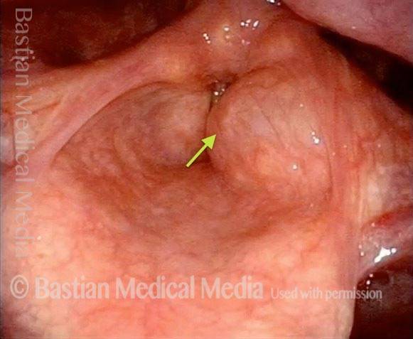
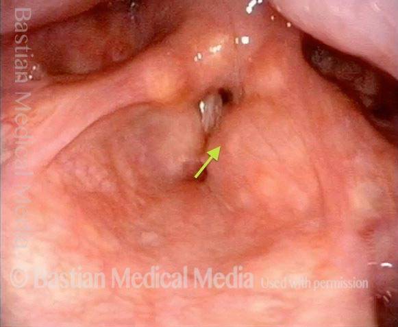
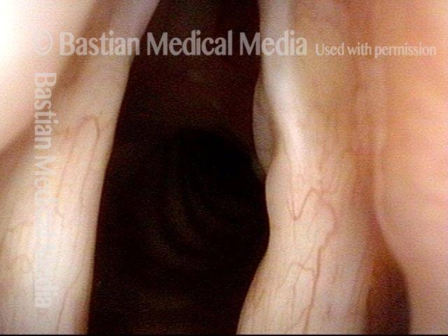
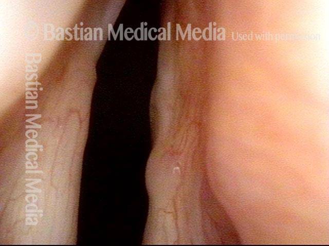
In some cases, the air that slips into and inflates the laryngocele will slip back out again as soon as the person stops making voice, so that the laryngocele abruptly inflates and deflates with each start and stop of speech or voice-making. (The photos and video below are an example of this.) In other cases, the air cannot exit as easily, but it may be reabsorbed slowly during quiet times or during sleep—only to be inflated again at the next instance of more active speaking.
Laryngocele vs. Saccular Cyst
A much more common disorder of the laryngeal saccule (compared with a laryngocele) is a saccular cyst, which can occur if the entrance to the laryngeal saccule becomes blocked. In this scenario, air is absorbed, but secretions build up and gradually expand the saccule.
Symptoms
A common symptom is hoarseness, because while the saccule is inflated, it may press press down on the vocal cords, not allowing them to vibrate freely, or it may block the laryngeal vestibule just above the cords and partially muffle the sound produced by the cords.
Treatment
Standard treatment is surgical removal, through one of two approaches: a small incision on the neck that leads into the larynx from the outside, or a laryngoscope that is inserted through the mouth and down into the larynx so that the laryngocele can be removed using a laser.
Photo Examples
Laryngocele (1 of 5)
Laryngocele (1 of 5)
Saccule (2 of 5)
Saccule (2 of 5)
Saccule blocks airway (3 of 5)
Saccule blocks airway (3 of 5)
Phonation ending (4 of 5)
Phonation ending (4 of 5)
Phonation ended (5 of 5)
Phonation ended (5 of 5)
Bilateral Laryngocele, Before and After Removal
Bilateral laryngocele (1 of 8)
Bilateral laryngocele (1 of 8)
Bilateral laryngocele (2 of 8)
Bilateral laryngocele (2 of 8)
Bilateral laryngocele (3 of 8)
Bilateral laryngocele (3 of 8)
Bilateral laryngocele (4 of 8)
Bilateral laryngocele (4 of 8)
Bilateral laryngocele, after removal (5 of 8)
Bilateral laryngocele, after removal (5 of 8)
Bilateral laryngocele, after removal (6 of 8)
Bilateral laryngocele, after removal (6 of 8)
Bilateral laryngocele, after removal (7 of 8)
Bilateral laryngocele, after removal (7 of 8)
Bilateral laryngocele, after removal (8 of 8)
Bilateral laryngocele, after removal (8 of 8)

Laryngocele: A Cause of Hoarseness
A laryngocele is a disorder of the saccule, or laryngeal appendix, in which air abnormally expands it. Watch this video to see how a laryngocele behaves in real-time, and why that can affect the voice.
